Autologous Oral Grafts

a technology of autologous and oral cavity, which is applied in the field of autologous oral grafts, can solve the problems of increasing sensitivity to heat and cold, exposing the root surface, and teeth may even loosen, so as to reduce the amount of inflamed or damaged oral mucosa tissue in the oral cavity of the patient, and reduce the amount of inflammation or damaged tissu

Inactive Publication Date: 2009-09-03
THE RES FOUND OF STATE UNIV OF NEW YORK
View PDF2 Cites 2 Cited by
  • Summary
  • Abstract
  • Description
  • Claims
  • Application Information

AI Technical Summary

Benefits of technology

[0009]In certain embodiments, the amount of inflamed or damaged oral mucosa tissue in the oral cavity of the patient is reduced as a result of being contacted with the therapeutic polypeptide (e.g. there is a 10%, or 20%, or 40%, or 50%, or 75% reduction in the amount of inflammation or damaged tissue in the oral cavity of the patient). In some embodiments, the biocompatible matrix comprises a biocompatible membrane. In particular embodiments, the oral mucosa tissue has been inflamed by infiltration of the oral mucosa with activated, maturing dendritic cells, including Langerhans cells (LC) and/or dermal dendritic cells (DDC) and T cells.
[0010]In certain embodiments, the cultured oral cells (e.g., oral keratinocytes) further comprise a second nucleic acid sequence encoding a reporter protein, wherein the reporter protein allows monitoring of expression of the therapeutic polypeptide. In particular embodiments, the reporter protein allows non-invasive monitoring of expression of the therapeutic polypeptide (e.g., e

Problems solved by technology

As that happens, the gums may recede, exposing the root surfaces and increasing sensitivity to heat and

Method used


Image

  • Autologous Oral Grafts
  • Autologous Oral Grafts
  • Autologous Oral Grafts

Examples


example 1

Generating Autologous Oral Grafts for Treating Oral Disease

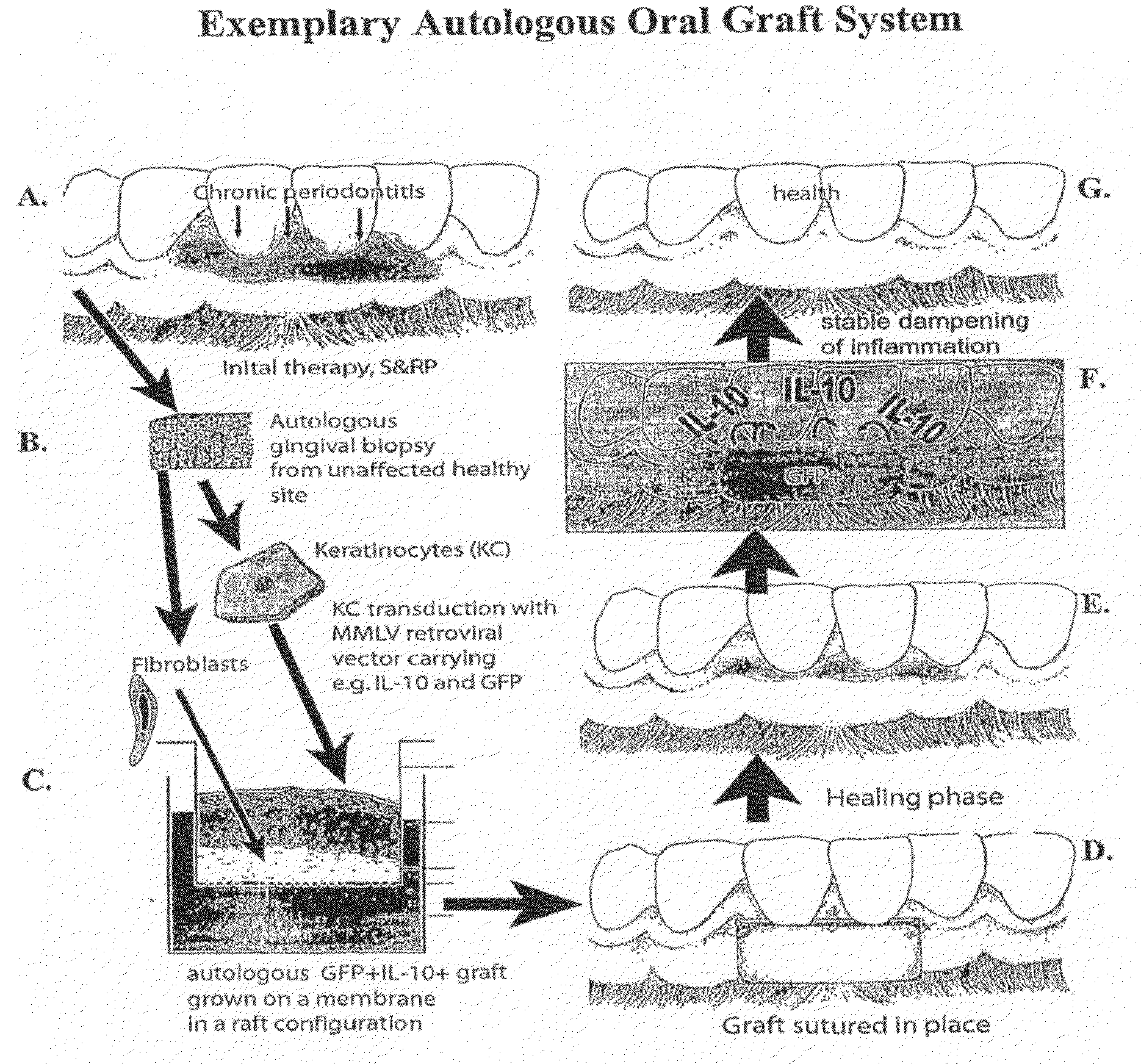
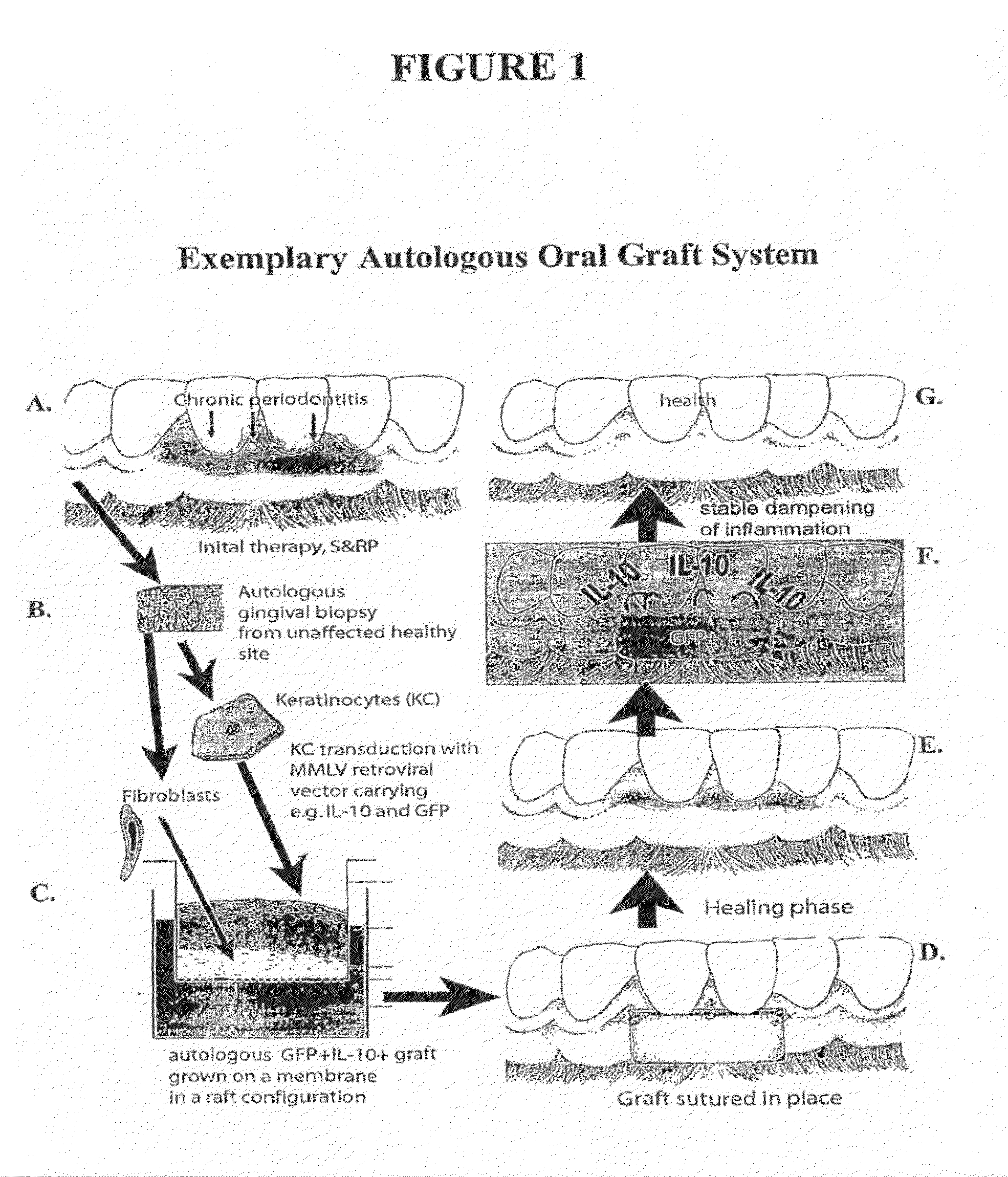
[0079]This Example describes the generation of an autologous oral graft for treating oral diseases, such as those involving tissue inflammation or damage. A subject with an oral disease such as chronic periodontitis, is identified as needing treatment. Under local anesthesia, a dental surgeon removes an autologous full-thickness oral mucosal biopsy (e.g., ˜5 mm pie-shaped wedge) of keratinized oral mucosa from a healthy oral mucosal site from the patient. A single resorbable suture is placed at the biopsy site. The oral mucosal biopsy is rinsed with antibiotic containing buffer, and immediately transferred aseptically into liquid nitrogen vial provided to the dentist by the clinical lab, and mailed overnight to a graft manufacturing facility.

[0080]Once at the graft manufacturing facility, the autologous oral mucosal tissue is thawed, diced and / or enzymatically digested through standard procedures, and keratinocytes (KC) and ...

example 2

Treating Oral Disease with an Autologous Oral Graft

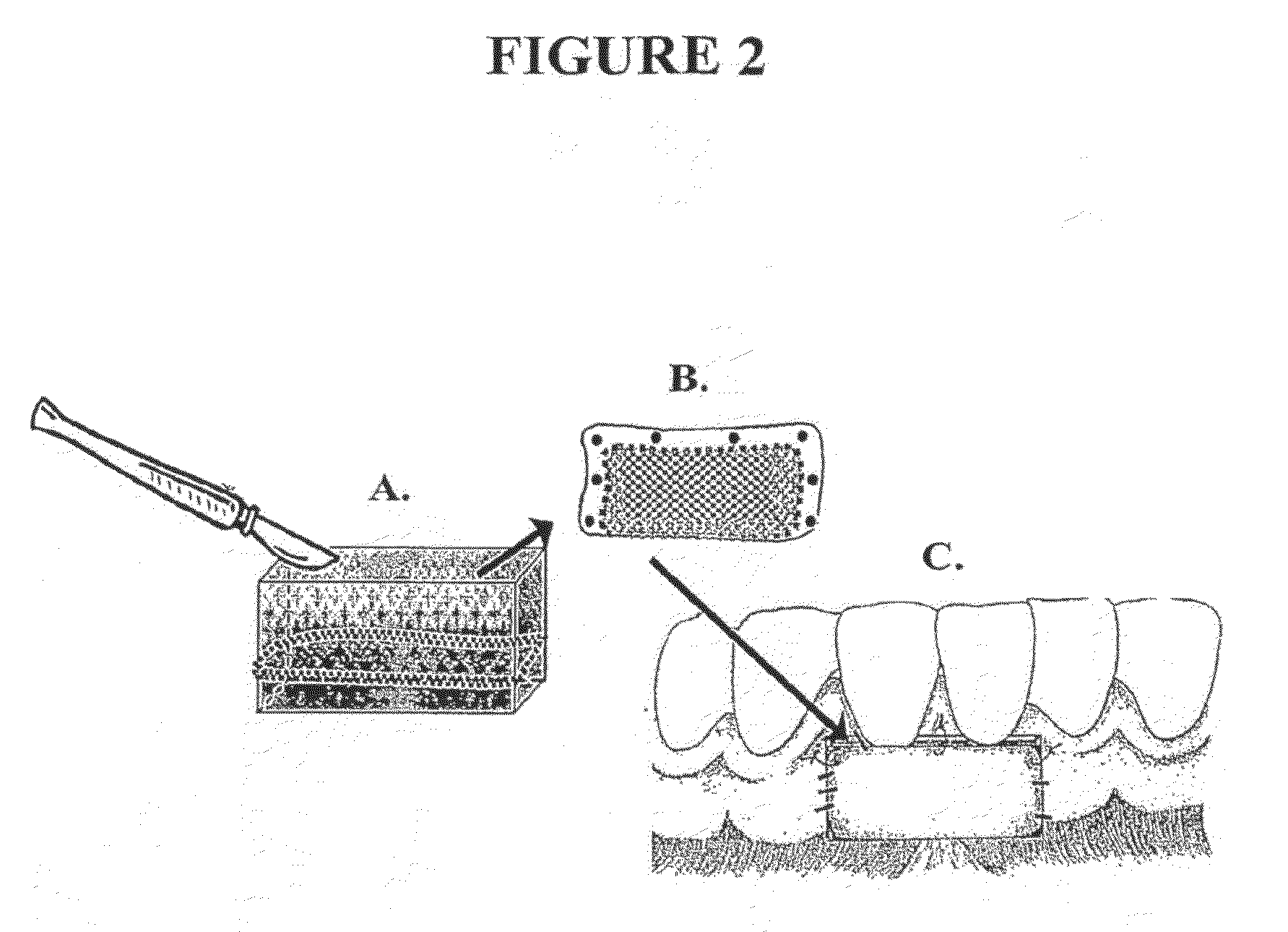
[0086]This Example describes the treatment of an oral disease with an autologous oral graft. In particular, a patient with chronic periodontitis is treated with the autologous oral graft prepared as described in Example 1. Initially, a surgeon prepares the graft recipient bed using a standard procedure for a free-oral mucosal graft. The autologous oral graft from Example 1 is removed from the vial by rimming it with a sterile 15 c scalpel while holding the oral graft with cotton forceps to prevent the graft from curling up prior to insertion. Next, the oral graft is inserted into the patient's mouth at the appropriate site along the gum tissue and is sutured in place with 5-0 gut chromic gut suture using a Castroviejo needle holder. Alternatively, the comers of the autologous oral graft are tacked down with surgical adhesive to stabilize the graft, and then it is sutured down tightly to the recipient bed. The autologous oral graft i...

example 3

Autologous Oral Grafts with Untransfected Oral Keratinocytes

[0088]In order to generate an autologous oral graft that employs untransfected oral keratinocytes, Example 1 could be repeated, while omitting the transfection steps. An oral graft generated in this manner could be employed to treat a patient with gingival recession, the result of damage to the oral mucosa by processes as described in Example 2. Treatment of oral disease with such un-transfected oral keratinocytes serves to help reduce the damage to the oral mucosa tissue or the oral calcified tissue in the oral cavity of the patient (e.g., by providing keratinized tissue and by secreting endogenous factors, such as cytokines and other proteins, that serve to reduce damage to the oral cavity).


PUM


Abstract

The present invention provides autologous oral grafts that may be used, for example, to treat oral cavity disease characterized by inflamed or damaged oral mucosa tissue, or to treat oral caries characterized by damaged enamel, dentin, or cementum. In certain embodiments, the autologous oral grafts comprise a biocompatible matrix and cultured oral cells (e.g., keratinocytes) from the patient to be treated. In certain embodiments, the cultured oral cells comprise an expression vector comprising a nucleic acid sequence encoding a therapeutic polypeptide configured to at least partially reduce the patient's oral mucosa tissue inflammation or damage. In particular embodiments, the oral disease is characterized by infiltration of the oral mucosa with activated, maturing dendritic cells.

Description

[0001]The present application claims priority to U.S. Provisional Application Ser. No. 60 / 798,816, filed May 9, 2006, which is herein incorporated by reference in its entirety.FIELD OF THE INVENTION[0002]The present invention relates to autologous oral grafts that may be used, for example, to treat oral cavity disease characterized by inflamed or damaged oral mucosa tissue, including periodontitis, mucogingival defects or gingival recession or to treat oral caries characterized by damaged enamel, dentin, or cementum. In certain embodiments, the autologous oral grafts comprise a biocompatible matrix and cultured oral cells (e.g., keratinocytes) from the patient to be treated. In certain embodiments, the cultured oral cells comprise an expression vector comprising a nucleic acid sequence encoding a therapeutic polypeptide configured to at least partially reduce the patient's oral mucosa tissue inflammation or damage. In particular embodiments, the oral disease is characterized by infi...

Claims


Application Information

Patent Timeline
IPC IPC(8): A61K9/14A61K45/00A61P1/02
CPCA61K35/12A61K48/0075A61L27/3804A61L27/3813A61L27/3839C12N2501/231A61L27/56A61L27/58C12N2510/00C12N2533/54C12N5/0632A61L27/3886A61P1/02
Inventor CUTLER, CHRISTOPHER W.
Owner THE RES FOUND OF STATE UNIV OF NEW YORK
Why Eureka
Try Eureka