

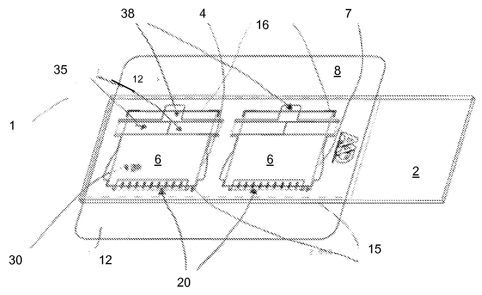
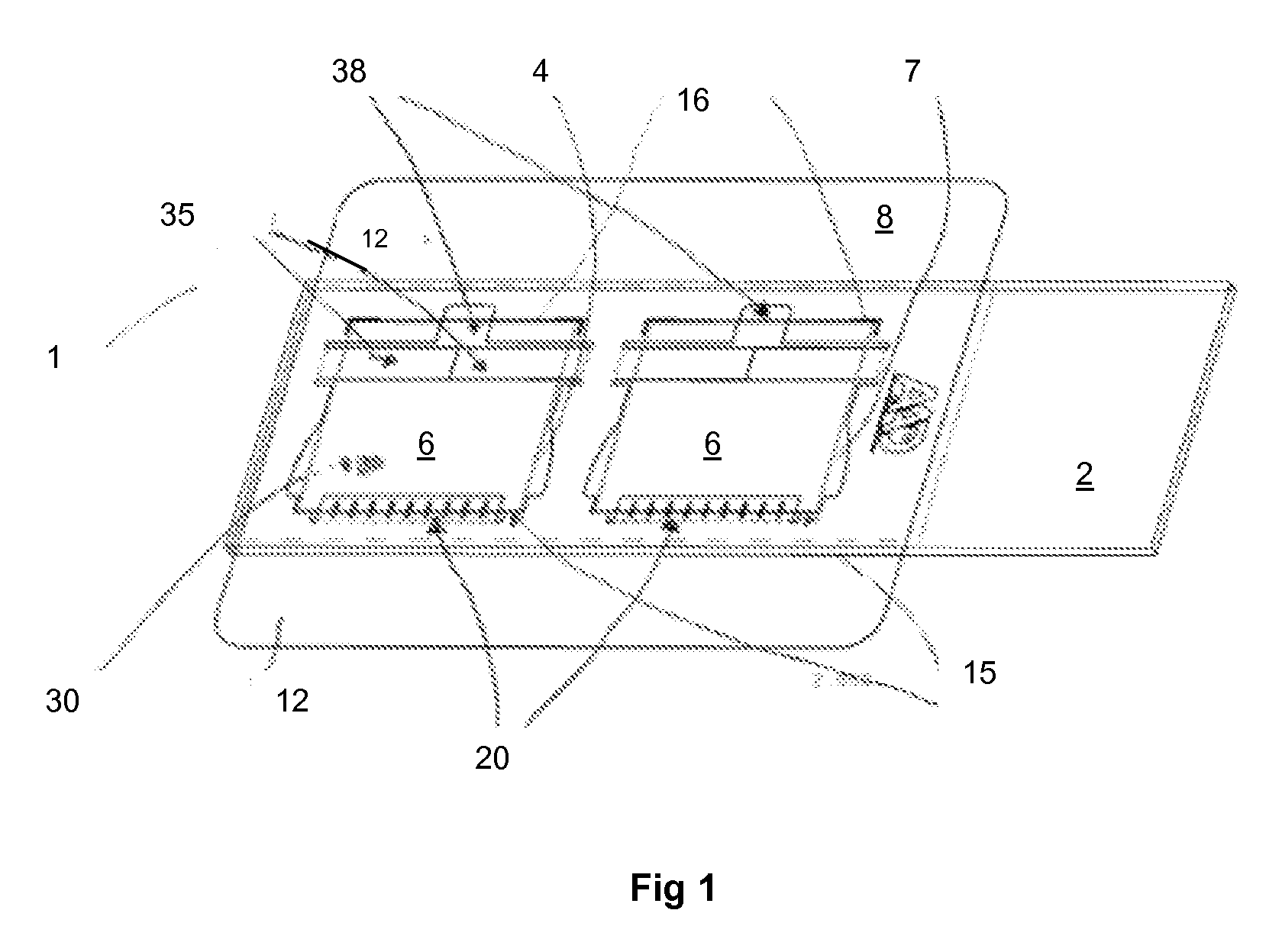
Microfluidic chamber assembly for mastitis assay
a microfluidic chamber and mastitis technology, applied in the field of microfluidic chambers, can solve the problems of increased labor costs, reduced sale value of culled cows, and early replacement of affected cows, and achieve the effect of small volume of milk
- Summary
- Abstract
- Description
- Claims
- Application Information
AI Technical Summary
Benefits of technology
Problems solved by technology
Method used
Image

Examples
example
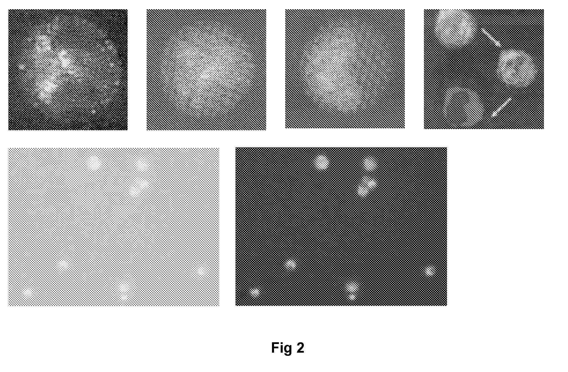
Assay for Milk Somatic Cell Differential Counts
[0042]In one embodiment of the assay, 80 μl of milk is mixed with 20 μl of a meta-chromatic stain, gently mixed, and a small drop of the mixture is placed in the deposition well of a slide of the invention. The wedge of the slide chamber fills automatically by capillary action, the cells in the milk are distributed evenly at optimum locations, and are ready for observation in seconds. A pre-concentration step may be required for very low SCC samples. The wedge can be aptly described as a “self preparing wet smear.”
[0043]Once the wedge slide has self-prepared, it is ready for immediate analysis by one of three methods:
(a) Visual identification by direct observation of the various live, intact, fluorescing cells, using a simple fluorescence microscope (for use by the experienced milk researcher); (b) Visual identification of the various cells using computer-enhanced digital camera images in a computer screen (for the use of a laboratory c...
PUM
| Property | Measurement | Unit |
|---|---|---|
| thickness | aaaaa | aaaaa |
| thickness | aaaaa | aaaaa |
| transparent | aaaaa | aaaaa |
Abstract
Description
Claims
Application Information