Electrosurgical system

a technology of electrosurgical and electrical burns, applied in the field of electrosurgical systems and methods, can solve the problems of electrical burns, bleeds, and the risk of certain types of patient injuries, and achieve the effect of increasing the functionality of the tool

Active Publication Date: 2009-10-01
APPL MEDICAL RESOURCES CORP
View PDF33 Cites 117 Cited by
  • Summary
  • Abstract
  • Description
  • Claims
  • Application Information

AI Technical Summary

Benefits of technology

[0019]At the same time, various embodiments of the surgical tools described herein can include different electrode configurations. I.e., while in one embodiment only the lower jaw is utilized to provide both coagulation and cutting functions, another embodiment can also employ the upper jaw element to be used in the coagulation and / or cutting process. In yet another embodiment, each jaw element can carry multiple electrode elements, greatly increasing the functionality of the tool. A specific electrode arrangement can allow for tools that are more suitable for particular surgical procedures.

Problems solved by technology

As bodily tissue, especially relatively highly vascularized tissue is cut during a surgical procedure, it tends to bleed.
Advantageously, unlike typical mechanical blade procedures, application of electrical energy to tissue tends to stop bleeding of the tissue.
Monopolar electrosurgical tools can be useful in certain procedures, but can include a risk of certain types of patient injuries such as electrical burns often at least partially attributable to functioning of the return electrode.
Even with the relatively focused surgical effects of bipolar electrosurgical tools, however, surgical outcomes are often highly dependent on surgeon skill.
For example, thermal tissue damage and necrosis can occur in instances where electrical energy is delivered for a relatively long duration or where a relatively high-powered electrical signal is delivered even for a short duration.
However, even for a highly experienced surgeon, it can be difficult for a surgeon to assess how quickly a mass of combined tissue types grasped in an electrosurgical instrument will be fused a desirable amount.
However, these systems have had shortcomings in that they have not provided consistent results at determining tissue coagulation, fusion, or cutting endpoints for varied tissue types or combined tissue masses.
These systems can also fail to provide consistent electrosurgical results among use of different tools having different tool and electrode geometries.
Typically, even where the change is a relatively minor upgrade to tool geometry during a product's lifespan, the electrosurgical generator must be recalibrated for each tool type to be used, a costly, time consuming procedure which can undesirably remove an electrosurgical generator from service.

Method used


Image

  • Electrosurgical system
  • Electrosurgical system
  • Electrosurgical system

Examples


Embodiment Construction

[0155]The following description is provided to enable any person skilled in the art to make and use the surgical tools and perform the methods described herein and sets forth the best modes contemplated by the inventors of carrying out their inventions. Various modifications, however, will remain apparent to those skilled in the art. It is contemplated that these modifications are within the scope of the present disclosure.

Electrosurgical System

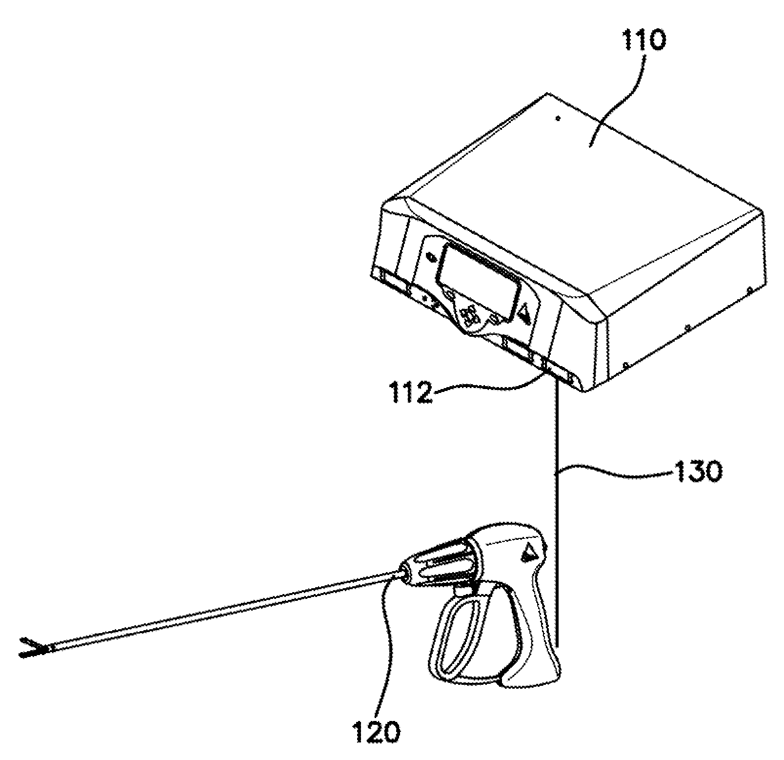
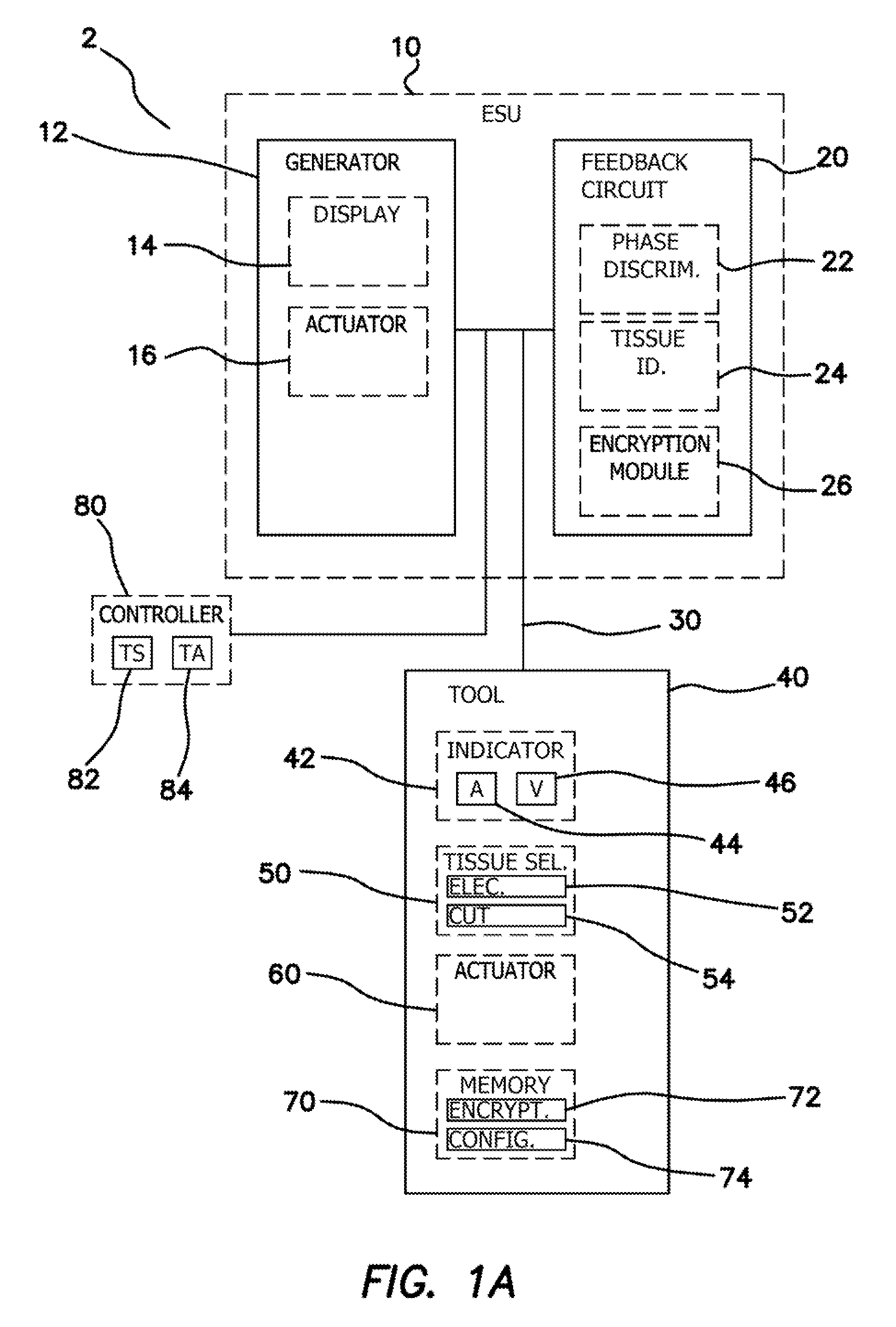
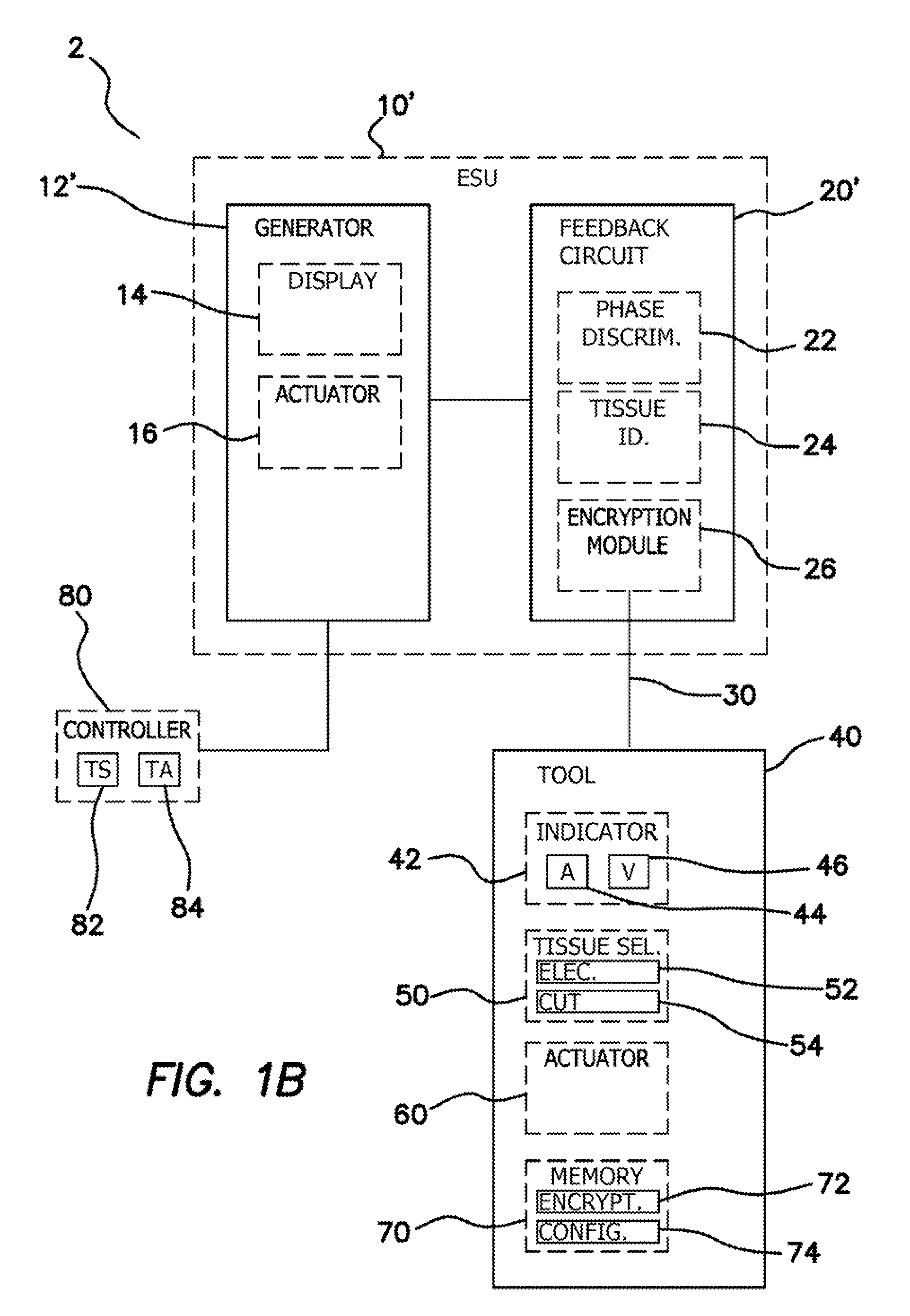
[0156]FIG. 1A illustrates a schematic diagram of an electrosurgical system 2. The electrosurgical system 2 can comprise an electrosurgical unit (ESU) 10 and an electrosurgical tool 40. The electrosurgical tool 40 can be electrically coupled to the electrosurgical unit 10. In some embodiments, an electronic coupler 30 such as an electrical wire, wire bundle, or cable can electrically couple the electrosurgical tool 40 to the ESU 10. In some embodiments, the electrosurgical system 2 can optionally further comprise an external tool controller 80...


PUM


Abstract

Phase end point determination is provided to automatically halt the application of energy to tissue. Prior to the application of energy, the phase end point determination is identified by measuring the product of permittivity and conductivity of the tissue to be treated.

Description

CROSS REFERENCE TO RELATED APPLICATIONS[0001]This application is a continuation of U.S. patent application Ser. No. 12 / 416,128, entitled “ELECTROSURGICAL SYSTEM”, filed Mar. 31, 2009, currently pending, which claims the benefit of U.S. Provisional Patent Application Ser. No. 61 / 040,980, entitled “FEEDBACK CONTROL MECHANISM FOR FUSING BIOLOGICAL TISSUE WITH HIGH FREQUENCY ELECTRICAL ENERGY”, filed Mar. 31, 2008; U.S. Provisional Patent Application Ser. No. 61 / 040,994, entitled “FUSING BIOLOGICAL TISSUE WITH HIGH FREQUENCY ELECTRICAL ENERGY”, filed Mar. 31, 2008; U.S. Provisional Patent Application Ser. No. 61 / 040,957, entitled “METHOD AND APPARATUS FOR BLOODLESS TISSUE DISSECTION”, filed Mar. 31, 2008; U.S. Provisional Patent Application Ser. No. 61 / 040,828, entitled “LAPAROSCOPIC BIPOLAR ELECTRICAL INSTRUMENT”, filed Mar. 31, 2008; U.S. Provisional Patent Application Ser. No. 61 / 040,890, entitled “APPARATUS AND METHOD FOR FUSION OF LIVING TISSUE”, filed Mar. 31, 2008; U.S. Provision...

Claims


Application Information

Patent Timeline
Patent Type & Authority Applications(United States)
IPC IPC(8): A61B18/18
CPCA61B18/1445A61B2018/00988A61B2017/2945A61B2018/00345A61B2018/00404A61B2018/00589A61B2018/00601A61B2018/00619A61B2018/00875A61B2018/124A61B2018/1412A61B2018/1432A61B2018/1455A61B18/1442A61B19/44A61B2018/00869A61B2018/00916A61B18/16A61B90/90A61B17/07207A61B18/1206A61B2017/00199A61B2018/00041A61B2018/00077A61B2018/00178A61B2018/00208A61B2018/00494A61B2018/00511A61B2018/00607A61B2018/00642A61B2018/00666A61B2018/00702A61B2018/00892A61B2018/00898A61B2018/00922A61B2018/126A61B2018/1266A61B2018/144A61B2018/1467A61B2018/162
Inventor FALKENSTEIN, ZORANCAPPELLO, CHRISTOPHER J.JOHNSON, GARY M.GIANNESCHI, BENJAMIN A.TRAN, OLIVIA J.WIXEY, MATTHEW A.PRAVONGVIENGKHAM, KENNIIPRAVONG, BOUNYAWATA, HARUYASUBECERRA, MATTHEW M.BRUSTAD, JOHN R.COHEN, ADAM J.HILAL, NABILPINGLETON, EDWARD D.HILAL, SAID S.HART, CHARLES C.WIKOFF, CHRIS R.
Owner APPL MEDICAL RESOURCES CORP
Why Eureka
Try Eureka