Methods of high rate, low profile tissue removal

a tissue removal and low-profile technology, applied in the field of tissue removal methods, systems and devices, can solve the problems of reproductive dysfunction, prolonged or heavy menstrual bleeding, pelvic pressure or pain, etc., and achieve the effect of reducing the risk of infection

Active Publication Date: 2009-10-29
HOLOGIC INC
View PDF99 Cites 129 Cited by
  • Summary
  • Abstract
  • Description
  • Claims
  • Application Information

AI Technical Summary

Benefits of technology

[0017]Additional aspects, features and advantages of the present invention will be set forth in part in the description which follows. The embodiments will be described in sufficient detail to enable those skilled in the art to practice the invention, and it is to be understood that other embodiments may be utilized and that structural or process changes may be made without departing from the scope of the invention. The following detailed description is, therefore, not to be taken in a limiting sense, and the scope of the present invention is best defined by the appended claims.

Problems solved by technology

In many instances, uterine fibroids can grow to be several centimeters in diameter and may cause symptoms like menorrhagia (prolonged or heavy menstrual bleeding), pelvic pressure or pain, and reproductive dysfunction.
However, current pharmacological therapies are largely ineffective and merely palliative.
However, despite their widespread use, hysterectomies also possess certain disadvantages, such as a loss of fertility, sexual dysfunction, and the risks commonly associated with a major surgical procedure, such as hemorrhaging, lesions, infections, pain and prolonged recovery.
Small particles are then injected from the catheter into the fibroid artery, blocking its blood supply and causing it to eventually shrink and die.
Although this procedure is less invasive than a hysterectomy, it often results in pain-related, post-surgical complications.
Moreover, the physicians that are trained to perform uterine artery embolization are typically interventional radiologists, as opposed to physicians trained specifically to take care of gynecological problems, whereas the physicians trained specifically to take care of gynecological problems typically do not possess the skill to perform catheter-based uterine artery embolization.
However, because the distending fluid is administered under pressure (which pressure may be as great as 100 mm Hg or greater), there is a risk, especially when tissue is cut, that the distending fluid may be taken up by a blood vessel in the uterus, i.e., intravasation, which uptake may be quite harmful to the patient.
As a result, due to the need for anesthesia, hysteroscopic resection is typically performed in a hospital operating room and, as a result, bears a large cost due to the setting and the support personnel required.

Method used


Image

  • Methods of high rate, low profile tissue removal
  • Methods of high rate, low profile tissue removal
  • Methods of high rate, low profile tissue removal

Examples


Embodiment Construction

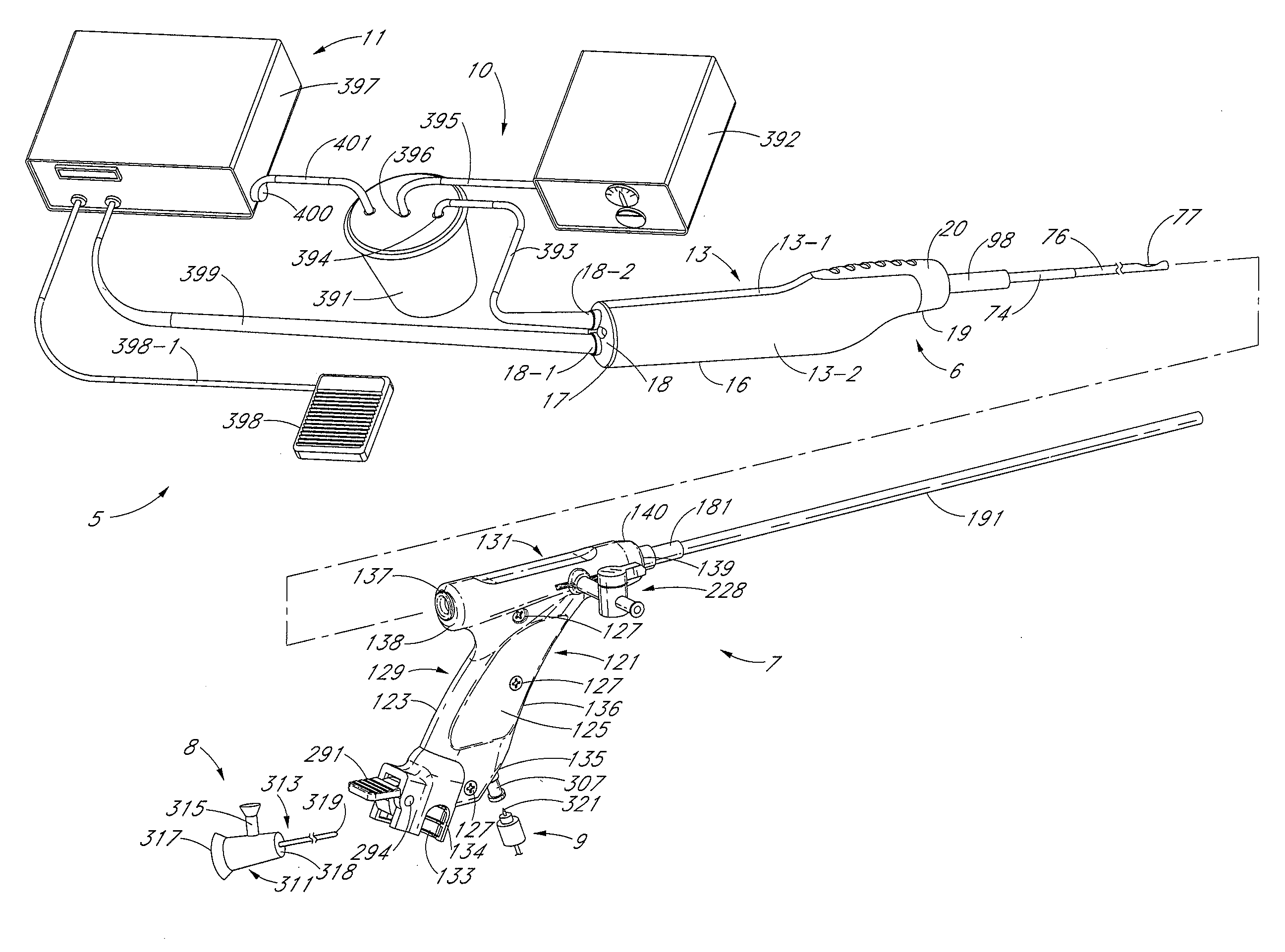
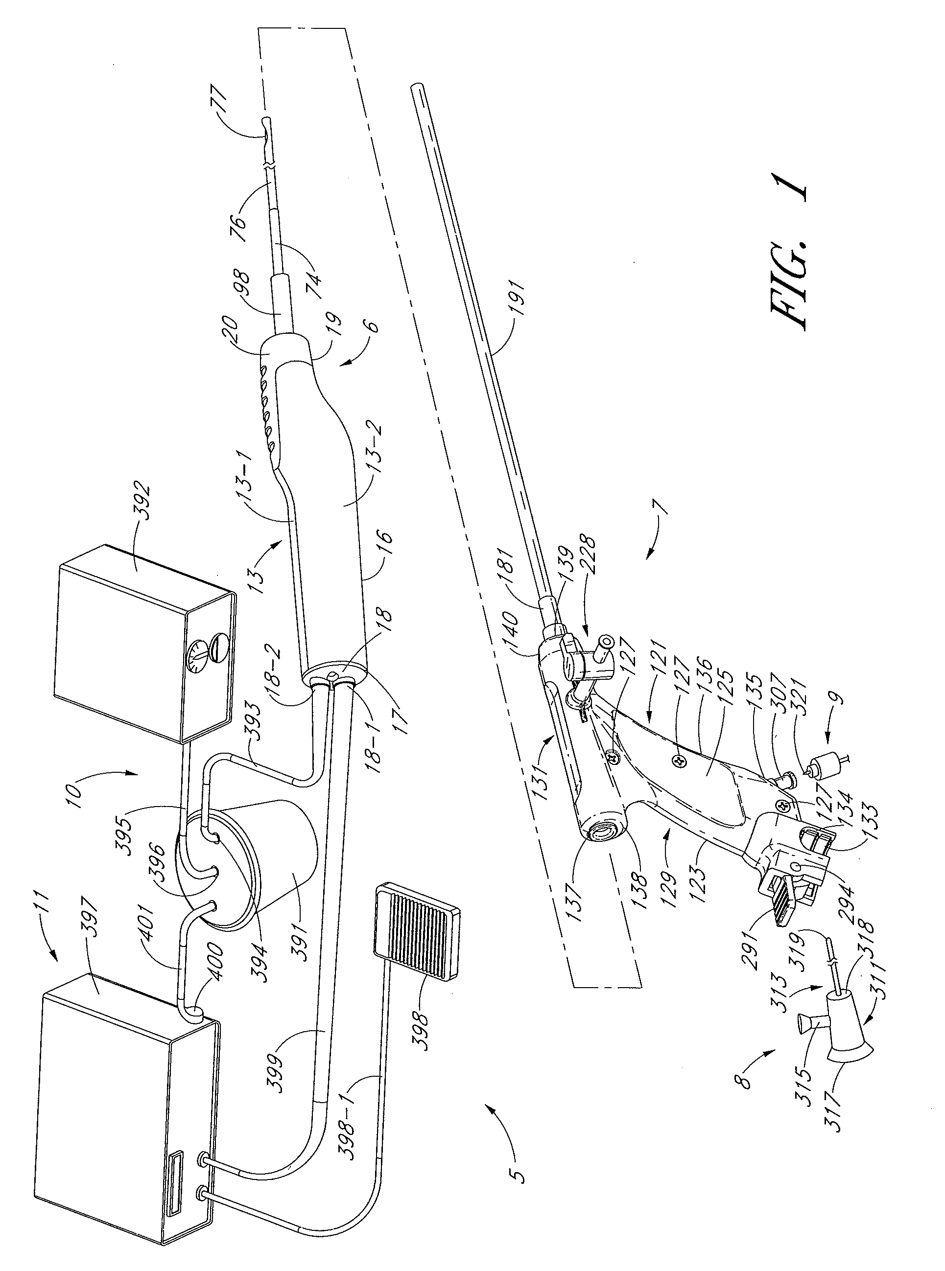
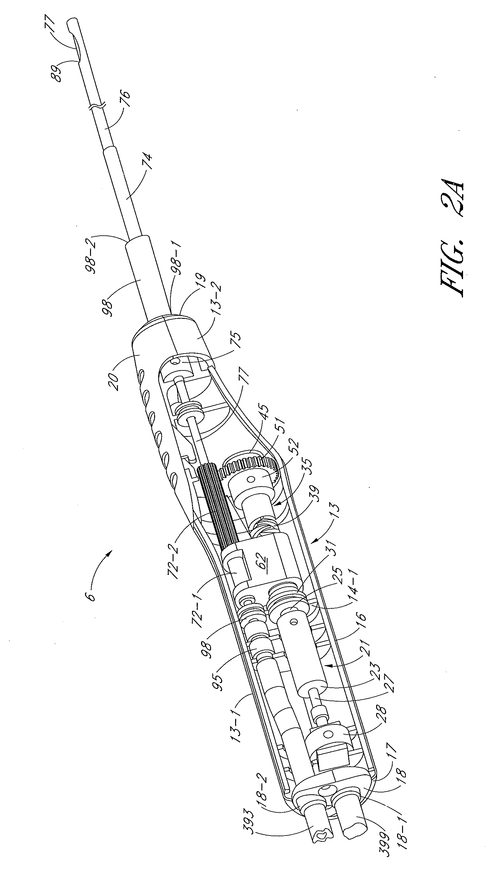
[0054]The present invention is described below primarily in the context of devices and procedures optimized for performing one or more therapeutic or diagnostic gynecological or urological procedures such as the removal of uterine fibroids or other abnormal uterine tissue. However, the devices and related procedures of the present invention may be used in a wide variety of applications throughout the body, through a variety of access pathways.

[0055]For example, the devices of the present invention can be optimized for use via open surgery, less invasive access such as laparoscopic access, or minimally invasive procedures such as via percutaneous access. In addition, the devices of the present invention can be configured for access to a therapeutic or diagnostic site via any of the body's natural openings to accomplish access via the ears, nose, mouth, and via trans-rectal, urethral and vaginal approach.

[0056]In addition to the performance of one or more gynecological and urologic pr...


PUM


Abstract

Disclosed are methods and devices for removing tissue from a site in a hollow organ, where the device has a low crossing profile and is capable of removing tissue at a high rate of speed. The device includes an elongate outer tube with a side opening and an inner tube moveably coaxially positioned within the outer tube. Tissue drawn into the side opening can be severed by moving the inner tube across the opening. Tissue may be removed through the device at a rate of at least about 1.4 cc per minute, through a lumen having a cross-sectional area of no greater than about 12.02 mm. Cutting may be accomplished by rotating the inner tube at a speed of at least about 4000 rpm, and axially reciprocating the inner tube at a rate of at least about 1.5 cycles per second. The window may have a rho value of no more than about 1, and the outside diameter of the device may be no more than about 3 mm.

Description

CROSS-REFERENCE TO RELATED APPLICATIONS[0001]The present application is a continuation in part of U.S. patent application Ser. No. 12 / 098,250, filed Apr. 4, 2008 (titled “METHOD, SYSTEM AND DEVICE FOR TISSUE REMOVAL”), which claims the benefit under 35 U.S.C. § 119(e) of U.S. Provisional Application 60 / 910,618, filed Apr. 6, 2007, U.S. Provisional Patent Application Ser. No. 60 / 910,625, filed Apr. 6, 2007, and U.S. Provisional Patent Application Ser. No. 60 / 986,912, filed Nov. 9, 2007 all of which are incorporated herein by reference. The present application is a continuation in part of U.S. patent application Ser. No. 12 / 098,318, filed Apr. 4, 2008 (titled “SYSTEM FOR USE IN PERFORMING A MEDICAL PROCEDURE AND INTRODUCER DEVICE SUITABLE FOR USE IN SAID SYSTEM”), which claims the benefit under 35 U.S.C. § 119(e) of U.S. Provisional Application Ser. No. 60 / 910,618, filed Apr. 6, 2007, and U.S. Provisional Patent Application Ser. No. 60 / 910,625, filed Apr. 6, 2007, all of which are inc...

Claims


Application Information

Patent Timeline
Patent Type & Authority Applications(United States)
IPC IPC(8): A61B17/32
CPCA61B17/29A61M1/0082A61B17/3201A61B17/32053A61B17/32056A61B17/320725A61B17/320783A61B17/3462A61B17/42A61B2017/00685A61B2017/00973A61B2017/2905A61B2017/320004A61B2017/320028A61B2017/320064A61B2017/320775A61B2017/3466A61B2017/4216A61B2018/1407A61B2217/005A61B2217/007A61B17/32002A61B1/303A61B17/3421A61B17/3496A61B17/3498A61B17/320758A61B8/12A61B2017/3445A61B2017/3447A61B2017/00331A61B2017/00738A61B2010/0208A61M1/842A61B90/30
Inventor ADAMS, RONALD DAVIDCHIN, ALBERT CHUN-CHISULLIVAN, ROY HEWITTLITSCHER, ERIC KARLCHURCHILL, WILLIAM LUCASGRUBER, WILLIAM HARWICK
Owner HOLOGIC INC
Why Eureka
Try Eureka