System and method for 3D vessel segmentation with minimal cuts

a technology of 3d vessel segmentation and minimal cuts, applied in image analysis, image enhancement, medical science, etc., can solve the problems of segmentation to “leak” into, challenge in vessel segmentation, and add a degree of difficulty in tracking the vessel path, etc., to achieve the effect of minimizing the energy of the form

Active Publication Date: 2009-11-12
SIEMENS HEALTHCARE GMBH
View PDF1 Cites 36 Cited by
  • Summary
  • Abstract
  • Description
  • Claims
  • Application Information

AI Technical Summary

Benefits of technology

[0020]According to a further aspect of the invention,

Problems solved by technology

Vessel segmentation is challenging, however.
First, branch points, such as bifurcations or trifurcations, add a degree of difficulty to tracking the vessel path.
Second, adjacent structures with similar signal intensities may cause a segmentation to “leak” into them.
Finally, intensity values within the vessel may vary due to uneven distribution of contrast or partial volume effects and can complicate segment

Method used


Image

  • System and method for 3D vessel segmentation with minimal cuts
  • System and method for 3D vessel segmentation with minimal cuts
  • System and method for 3D vessel segmentation with minimal cuts

Examples


Embodiment Construction

[0039]Exemplary embodiments of the invention as described herein generally include systems and methods for segmentation of vascular structures in volumetric image datasets. Accordingly, while the invention is susceptible to various modifications and alternative forms, specific embodiments thereof are shown by way of example in the drawings and will herein be described in detail. It should be understood, however, that there is no intent to limit the invention to the particular forms disclosed, but on the contrary, the invention is to cover all modifications, equivalents, and alternatives falling within the spirit and scope of the invention.

[0040]As used herein, the term “image” refers to multi-dimensional data composed of discrete image elements (e.g., pixels for 2-D images and voxels for 3-D images). The image may be, for example, a medical image of a subject collected by computer tomography, magnetic resonance imaging, ultrasound, or any other medical imaging system known to one of...


PUM


Abstract

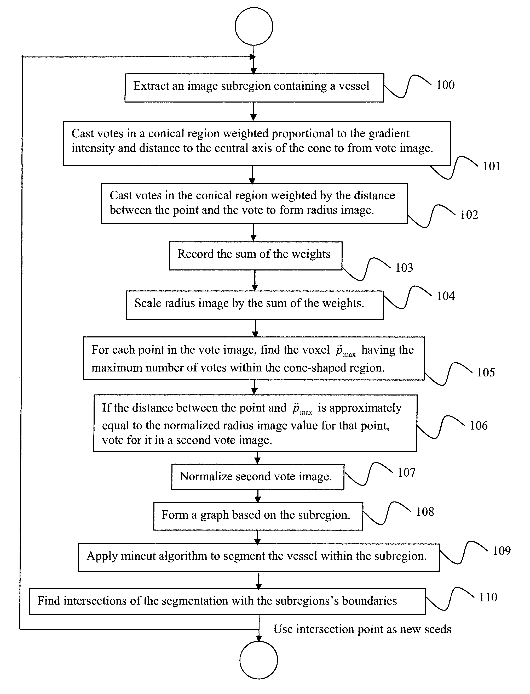
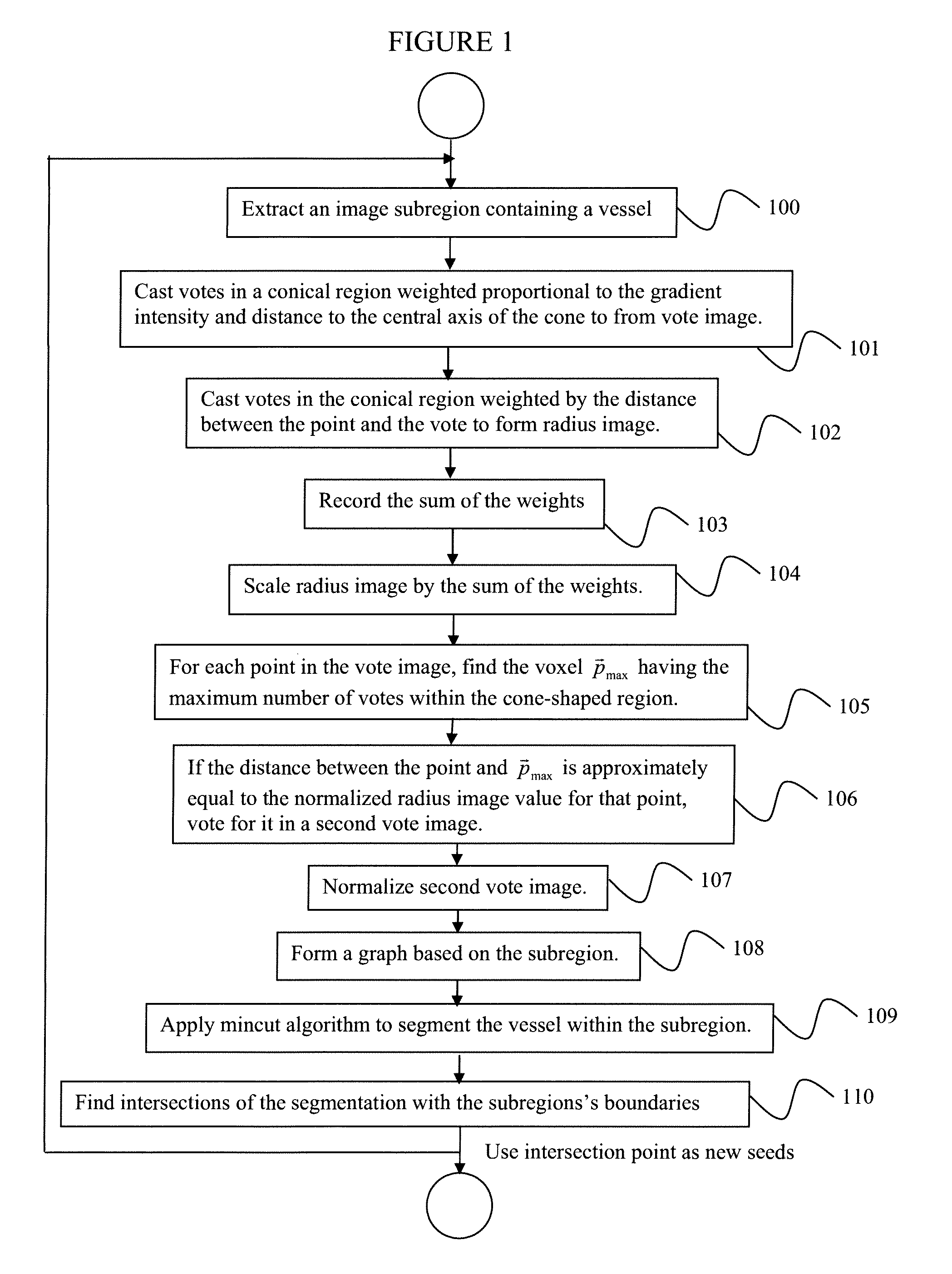
A method for segmenting tubular structures in digital medical images includes extracting a subregion from a 3-dimensional (3D) digital image volume containing a vessel of interest, identifying potential vessel centerpoints for each voxel in the subregion by attaching to each voxel a tip of a 3D cone that is oriented in the direction of the voxel's image gradient and having each voxel within the cone vote for those voxels most likely to belong to a vessel centerline, selecting candidates for a second vote image that are both popular according to a first vote image, as well as being consistently voted upon by a radius image, reconfiguring the subregion as a graph where each voxel is represented by a node that is connected to 26 nearest neighbors by n-link edges, and applying a min-cut algorithm to segment the vessel within the subregion.

Description

CROSS REFERENCE TO RELATED UNITED STATES APPLICATIONS[0001]This application claims priority from U.S. Provisional Application No. 61 / 033,958 of Dikici, et al., filed Mar. 5, 2008, and from U.S. Provisional Application No. 61 / 036,607 of Dikici, et al., filed Mar. 14, 2008, the contents of both of which are herein incorporated by reference in their entireties.TECHNICAL FIELD[0002]This disclosure is directed to the segmentation of vascular structures in volumetric image datasets.DISCUSSION OF THE RELATED ART[0003]Segmentation of vascular structures in volumetric datasets facilitates several forms of useful analysis. For example, quantification of the dimensions of an aortic aneurysm helps to determine the time point for intervention. In coronary artery disease, the degree of latency of a stenotic lesion influences treatment decisions. And, the planning for the placement of stents and by-pass graphs benefits greatly from the delineation of the vessel boundaries. Finally, a by-product of...

Claims


Application Information

Patent Timeline
IPC IPC(8): G06K9/34
CPCG06T7/0081G06T7/0093G06T2207/30101G06T2207/20044G06T2207/20168G06T2207/10072Y10S128/922G06T7/11G06T7/162
Inventor DIKICI, ENGINO'DONNELL, THOMAS P.SETSER, RANDOLPH M.WHITE, RICHARD D.
Owner SIEMENS HEALTHCARE GMBH
Why Eureka
Try Eureka