Small-scale diagnosis system

a diagnostic system and small-scale technology, applied in the field of small-scale diagnostic systems, can solve problems such as mismatching of patient and image data, and achieve the effects of relieved doctor's burden, easy sorted photographic data, and convenient sorted photographic data

Inactive Publication Date: 2009-11-12
KONICA MINOLTA MEDICAL & GRAPHICS INC
View PDF5 Cites 19 Cited by
  • Summary
  • Abstract
  • Description
  • Claims
  • Application Information

AI Technical Summary

Benefits of technology

[0023]According to the invention described in claim 1, operations such as creation of a defined photographic image data, matching of the defined photographic image data with examination object information and displaying photographic image data had been conducted in individual device such as the console and the viewer can be conducted in one control device. Therefore, in a circumstance of a relatively small-scale facility such as private medical practice where staff and facility are few, there can be an effect that the burden of the doctor can be relieved, since the plurality of devices are not necessary to be operated.
[0024]That is, in a circumstance where the number of doctors and assistants to operate various kinds of devices are few, or one doctor conducts from photographing to medical examination in some occasion, there is less possibility of mismatching the patient and the photographic data. Thus in the invention described in claim 1, for example, a control device such as a workstation (PC) where a console to operate image forming apparatus and a viewer to display the photographic data for observation are combined is provided in a room where the doctor to conducts medical examination is installed. After photographing the patient, the image data is immediately displayed on the control device and image processing such as tone processing is carried out if needed, thereafter the doctor conducts medical examination based on the photographic data. Thereby duplicated input operation through key board by the doctor, a nurse or the assistant, and setting and operation of the plurality of the devices are not necessary. Thus the doctor can concentrate medical examination.
[0025]Also, since correlating of the defined photographic data representing a final image for examination with the patient can be conducted by the same control device, photographic data can be sorted out readily without operating special devices or inputting photographic image order information. Thus the burden of the doctor can be relieved. Further, as above, after correlating the photographic data with information related to the examination object such as a name of the patient, the photographic data is stored in a storing device such as a server, therefore, the photographic image data can be effectively utilized as a comparison image to examine progress of curing for next examination.
[0026]Furthermore, since the photographic image data is displayed on the display device for examination, a film is not used to store the photographic image data and examination data. Therefore a cost is saved.
[0027]According to the invention described in claim 2, with a plurality of the image forming apparatuses, photographing of a plurality of the patients can be conducted simultaneously. Also, in case photographing is conducted using the plurality of image forming apparatuses, the created photographic data is transmitted to one control device such as the workstation (PC) installed, for example, in a room where the doctor conducts examination, and processed. Thus operator such as the doctor can sort the photographic data readily without operating special devices or a keyboard for input. Thus, in the small-scale facility where staff such as doctor is few, there is provided an effect to improve a working environment by relieve the burden of the doctor so that the doctor can concentrate on medical examination.
[0028]According to the invention described in claim 3, since photographing can be conducted through a plurality of kinds of image forming apparatus such as radiographic imager, ultrasonograph and endoscope imager, an appropriate apparatus for each patient can be used for photographing in accordance with requirement. In this case, the created photographic image data is transmitted to one control device such as the workstation (PC) installed, for example, in the room where the doctor conducts examinations and processed. Thus operator such as the doctor can sort the photographic image data readily without operating special devices or a keyboard for input. Thus, in the small-scale facility where staff such as doctor is few, there is provided an effect to improve a working environment by relieve the burden of the doctor so that the doctor can concentrate on medical examination.

Problems solved by technology

Therefore, there is a risk that patients and image data are mismatched.

Method used


Image

  • Small-scale diagnosis system
  • Small-scale diagnosis system
  • Small-scale diagnosis system

Examples


first embodiment

[0079]First, a first embodiment of a small-scale medical diagnosis system related to the present invention will be described with reference to the FIG. 1 to FIG. 10.

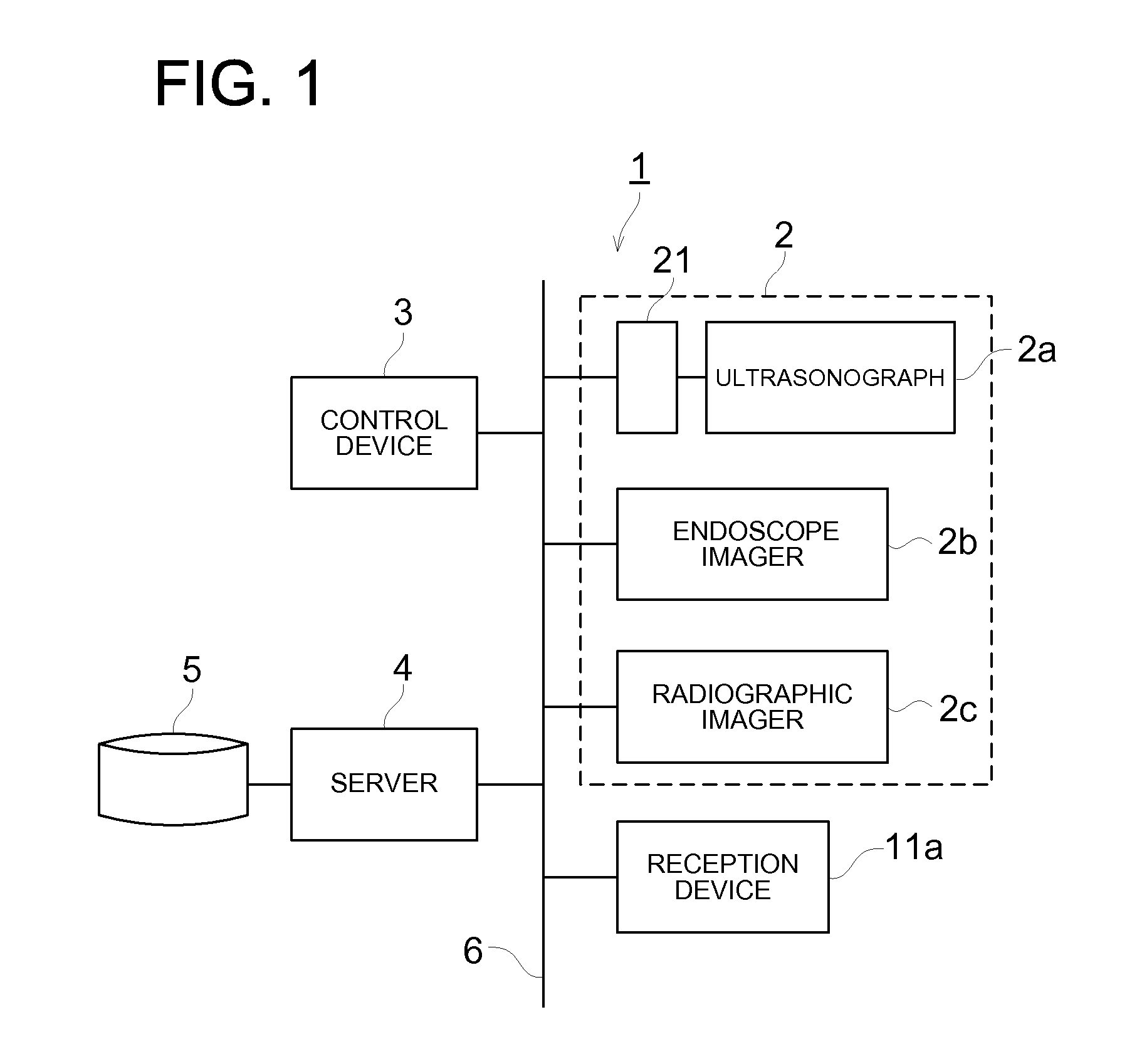
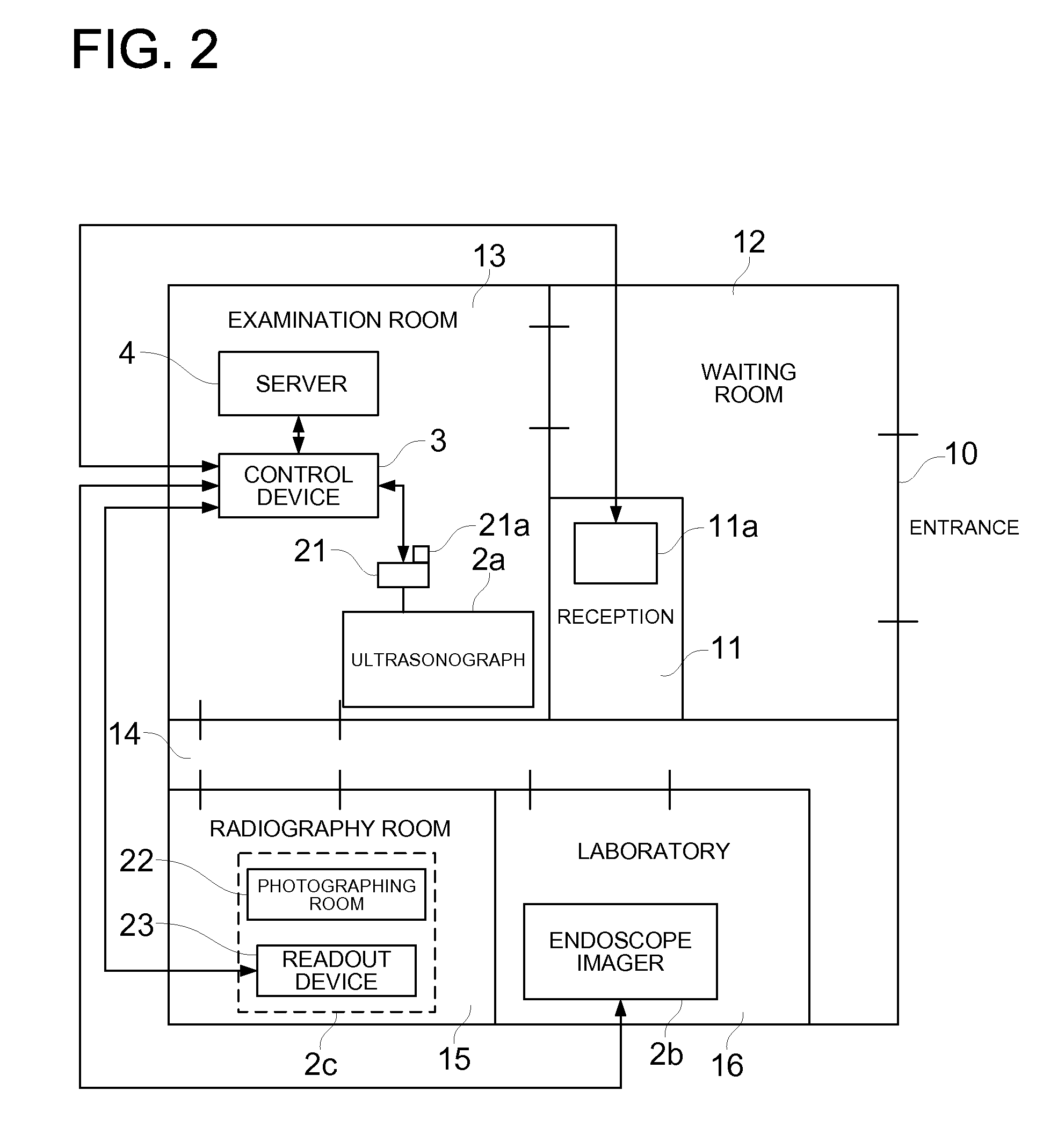
[0080]FIG. 1 is shows a system configuration of a small-scale medical diagnosis system 1 related to the present embodiment, and FIG. 2 shows an exemplary lay-out of each apparatus in a medical facility in case the small-scale medical diagnosis system 1 is utilized.

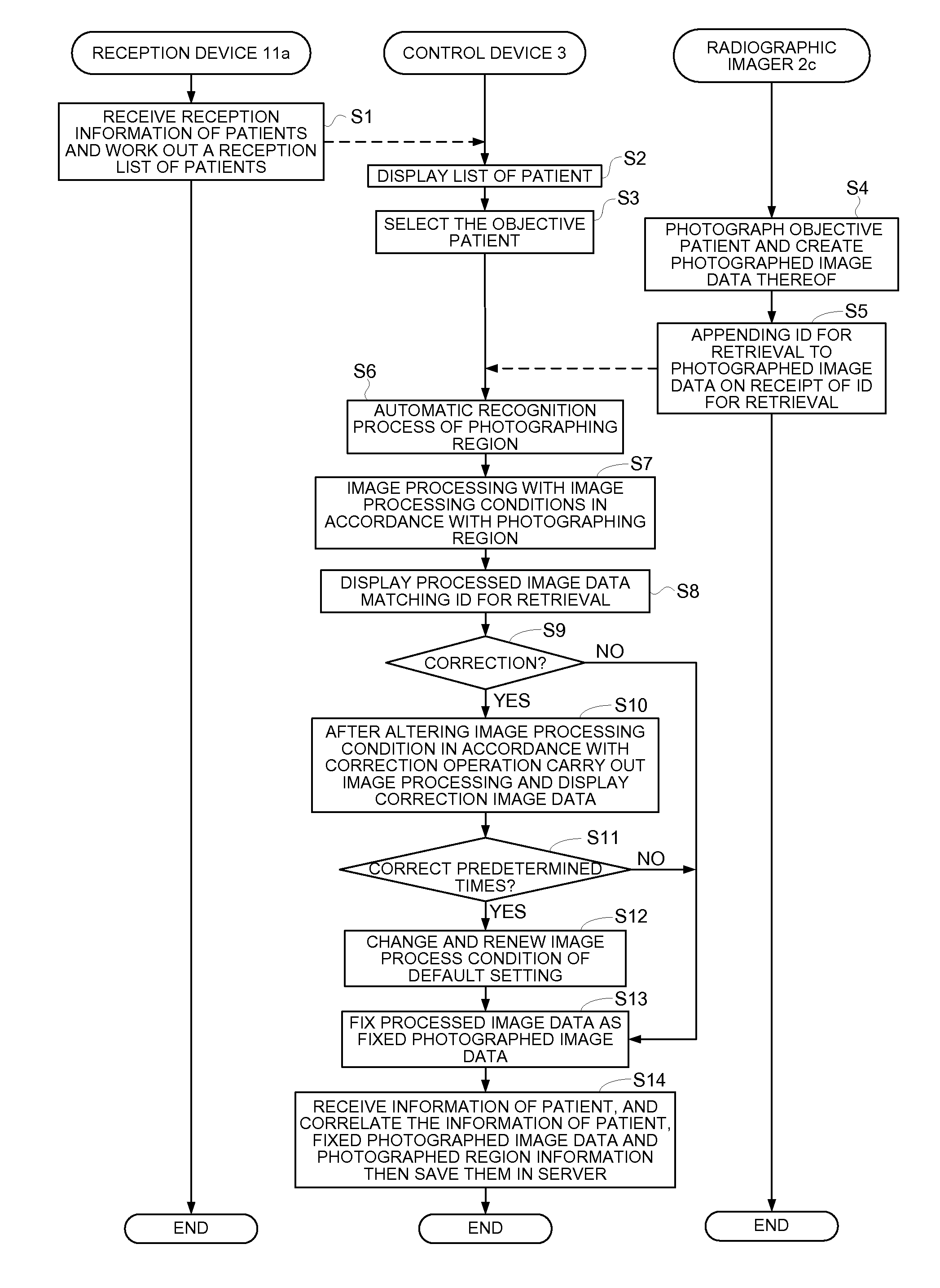
[0081]The small-scale medical diagnosis system is a system to carry out a successive operation from reception of a patient, to medical examination of a doctor and payment, which is applied to relatively small-scale medical facilities such as a medical practice or a clinic. As FIG. 1 shows, the small-scale medical diagnosis system 1 is configured with an ultrasonograph 2a, endoscope imager 2b and radiographic imager 2c which represent image creating apparatus 2, a control device 3, a server 4 and a reception device 11a, and each of the apparatuses and devices is...

second embodiment

[0181]Next, a second embodiment of the small-scale diagnosis system related to the present invention will be described. In the first embodiment described above, to recognize the photographed region of the photographic image, by analyzing the photographic image data from scratch, the photographed region is automatically recognized, however in the present embodiment, using a human body region icon displayed on the display section, the doctor select a broad photographing region, and the photographed region is automatically recognized based on information of the broad region.

[0182]Meanwhile, the small-scale diagnosis system 1 in the present embodiment has the same configuration as the first embodiment described above, and the portions having been described are denoted by the same symbol to omit the description.

[0183]In the following description, as the radiographic imager 2c of the image creating apparatus 2, a CR device using a portable cassette in which a photostimulable phosphor plat...


PUM


Abstract

The present invention is to provide a small-scale diagnosis system for conducting simple and efficient examination, including: an image creating apparatus for creating photographic image data of examination object; a CPU to conduct image processing for creating defined photographic image data from the photographic image data; an input section to input the examination object information to specify the photographed examination object; a CPU to correlate the defined image data created and examination object information corresponding to the defined photographic image data; a server to store the defined photographic image data having been correlated and the examination object information; a display section capable of displaying at least any one of the photographic image data, the defined photographic image data or the examination object information; and a control device having at least the CPU to carry out image processing, the CPU to carry out correlation processing and the display section.

Description

FIELD OF THE INVENTION[0001]The present invention relates to a small-scale diagnosis system, and in particular, to a small-scale diagnosis system used in small-scale medical facility.PRIOR ART[0002]There has been know an diagnosis system where using image creating forming apparatuses such as a CR (Computed Radiography) device or FPD (Flat Panel Detector), a technician photographs a patient who is a object of examination and carries out image processing such as tone processing to make the obtained image suitable for examination, then outputs the image having been processed by image processing on a film to be made available for a doctor to carry out interpretation of radiogram.[0003]With such diagnosis system, a plurality of persons shear roles to carry out examination, namely a person (receptionist) to receive a patient visited a hospital, a person (technician) to actually photograph the patient in a photographing room and creates a digital image, a person (a technician who is assign...

Claims


Application Information

Patent Timeline
Patent Type & Authority Applications(United States)
IPC IPC(8): G06T7/00
CPCA61B1/04A61B6/00A61B6/463G06T2207/30004A61B8/00A61B8/4416G06T7/0012A61B6/467
Inventor KAJI, DAISUKEUMEKI, MAMORUYONEKAWA, HISASHIOKUZAWA, JIROMOTOKI, WATARUSHIIBASHI, TAKAOMURAOKA, SHINTARO
Owner KONICA MINOLTA MEDICAL & GRAPHICS INC
Why Eureka
Try Eureka