Biosensors for monitoring receptor-mediated g-protein activation

a biosensor and receptor technology, applied in the field of biosensors, can solve the problems of largely in vitro studies deduced from classical collision models, unable to direct assess the real-time interactions between receptors and g proteins in living cells, and the general acceptance of collision-based models

Active Publication Date: 2009-12-03
UNIV DE MONTREAL
View PDF1 Cites 17 Cited by
  • Summary
  • Abstract
  • Description
  • Claims
  • Application Information

AI Technical Summary

Benefits of technology

[0008]In recent years, fluorescence and bioluminescence resonance energy transfer approaches (FRET and BRET) have been increasingly used to study protein-protein interactions and appreciate dynamic changes in protein conformation16, 17. The dependence of the energy transfer efficacy on the distance between energy donors and acceptors permits real time measurements that are both sensitive and specific to the labelling sites of the proteins thus allowing inference on the dynamic structural changes18-22.
[0010]In addition, the invention provides a useful tool to probe for conformational changes occurring in the receptor / G protein complexes and G protein heterotrimer resulting from ligand binding to the receptor. As a result, by multiplexing different BRET-biosensors of the receptor / G protein or the G protein complex itself, the invention offers the possibility to set up pharmacological fingerprints that are specific to each receptor ligand, thus allowing to differentiate the distinct signalling modes of different ligand toward the various signalling pathways engaged.
[0011]Results demonstrate that GPCR fused to the energy donor Renilla luciferase (RLuc) and G protein subunits attached to a green fluorescent protein (YFP, GFP10 or GFP2) energy acceptor or vice versa, provide reliable biosensors that can directly monitor the kinetic and selectivity of G protein engagement upon receptor activation. In addition to offering a new generally applicable method to probe dynamic protein interactions involved in G protein activation, the approach offers an advantageous tool to monitor the activation of distinct G protein subunits using a single assay mode. Receptor-mediated G protein activation can also be detected through a biosensor using the Gβ or Gγ subunits fused to RLuc and the Gα subunit fused to GFP10, GFP2 or YFP and / or vice versa, thus allowing to monitor interactions between Gα and Gβ or Gγ subunits fusion proteins as a result of activation of an untagged receptor. The data described herein provides direct kinetic measurement of the receptor-mediated activation of heterotrimeric G proteins in living cells, allowing the detection of both activation and desensitization events.
[0012]Due to its sensitivity and applicability for all major classes of Gα subunits (Gαs, Gαi / o, Gαq / 11, Gα12 / 13), the receptor / Gβγ interaction BRET assay can be viewed as general method to monitor receptor-mediated G protein activation. In that respect, it may be considered superior to the widely used GTPγS binding assay since the nucleotide binding method has proven difficult for Gs and Gq coupled receptor due to the very weak signal / noise ratio obtained with these Gα subunits. When compared with the classical second messenger-based read-out that are classically used to identify the G proteins that can be activated by a given receptor, the BRET-based assays present the advantage of a single and homogeneous assay mode that directly measure the engagement of the G protein. Thus the receptor / G protein interaction BRET assay could advantageously be used as a general strategy for the identification of ligands for GPCRs.

Problems solved by technology

However, our knowledge concerning the molecular events determining receptor signaling efficacy remains rudimentary.
However, this generally-accepted classical collision-based model, deduced largely from in vitro studies, has recently been challenged.
At present, no method permits the direct assessment of the real-time interactions between receptors and G protein in living cells.

Method used


Image

  • Biosensors for monitoring receptor-mediated g-protein activation
  • Biosensors for monitoring receptor-mediated g-protein activation
  • Biosensors for monitoring receptor-mediated g-protein activation

Examples


example 1

Configuration of the BRET Partners Used to Monitor Receptor-Mediated G Protein Activation

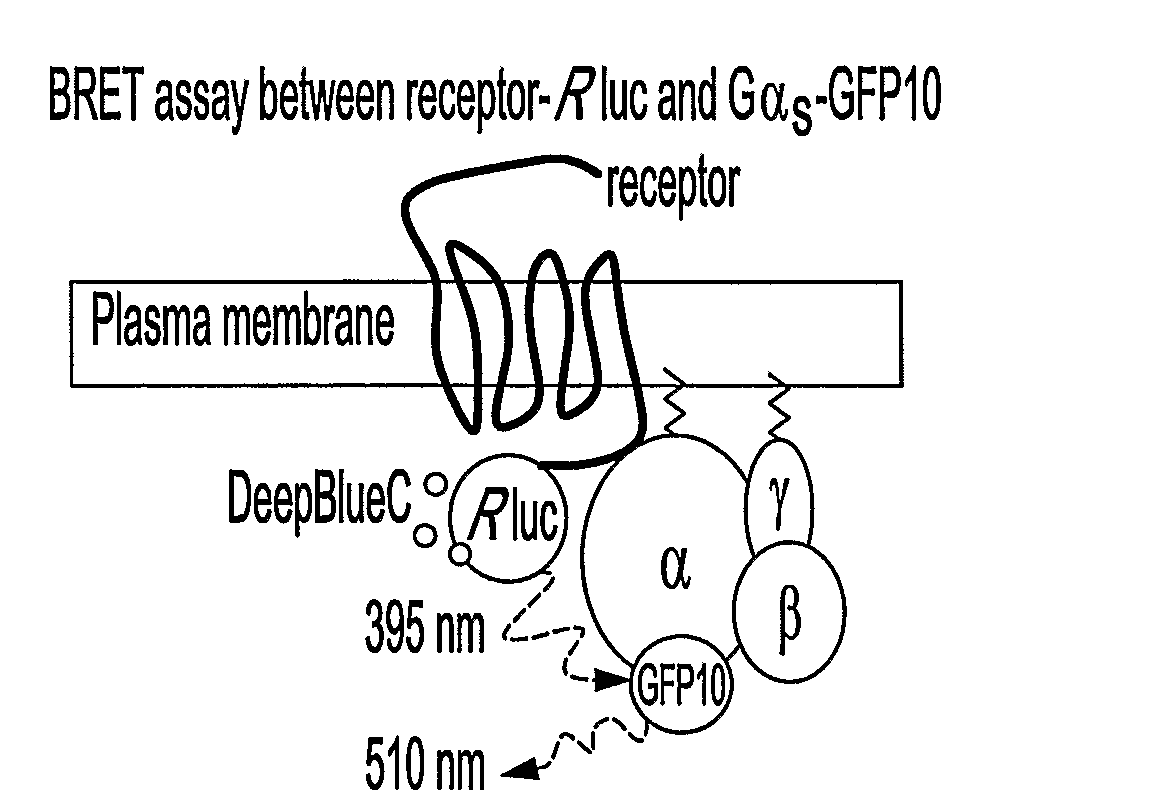
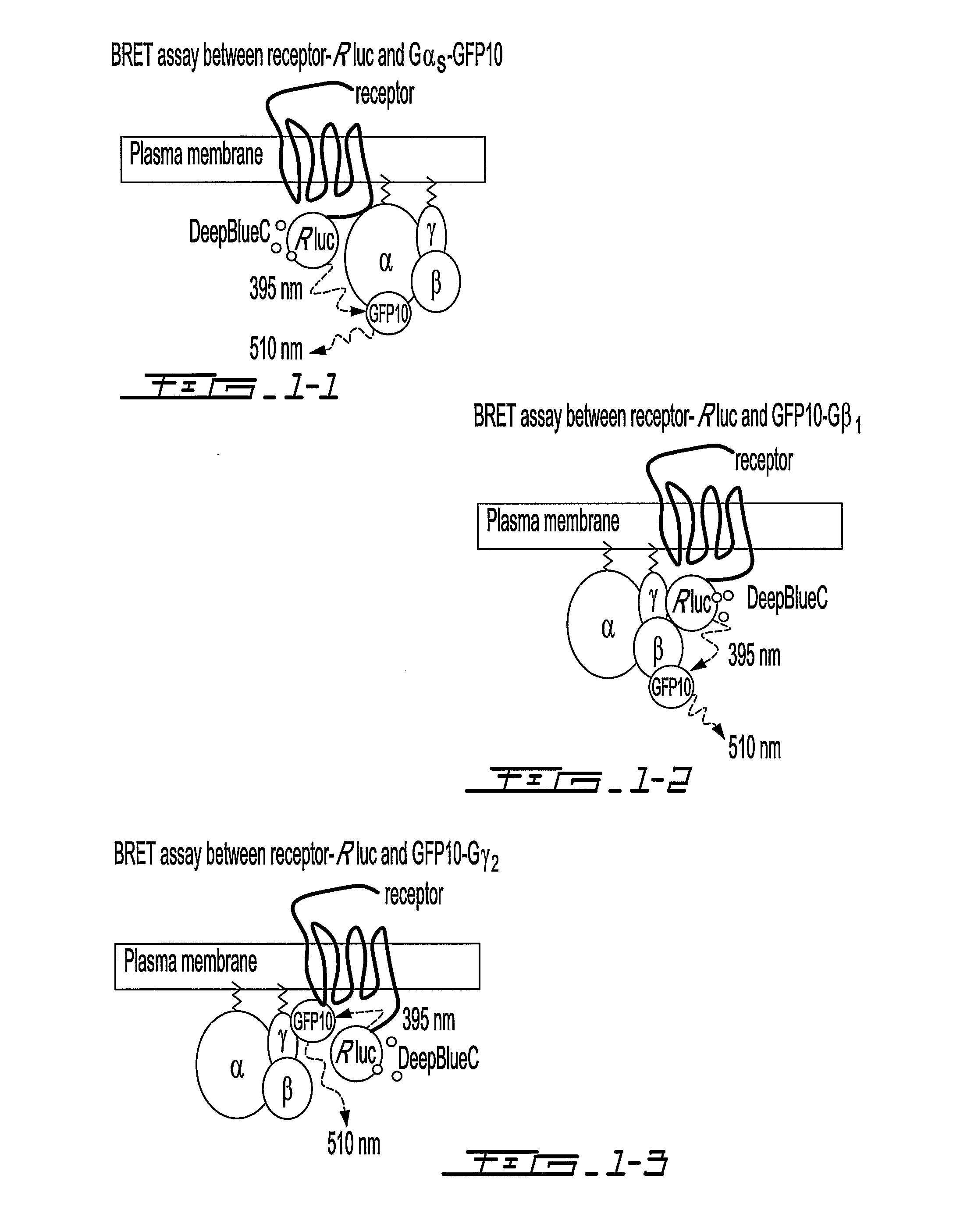
[0112]To monitor the interactions between GPCRs and their cognate G proteins and among the G proteins subunits, BRET1 and BRET2 signals were measured between the different partners in living cells in the presence and absence of selective receptor ligands. Receptors, Gαi1, Gβ1 and Gγ2 were fused to BRET energy donor or acceptor (FIG. 15a). The receptors (α2AR, β2AR and CRLR) were fused at their carboxyl tail to the Renilla reniformis luciferase (Rluc) or Aequorea victoria green fluorescent proteins (GFP2, GFP10 or YFP, depending on the partners considered). Gβ1 and Gγ2 were fused to their N-terminus to either Rluc or GFP10. For Gαi1, two different constructs were engineered so as to allow a better monitoring of the relative movements of the partners. Rluc was inserted in connecting loops located at opposite sides within the helical domain of the protein (FIG. 15b). One of the insertion site betwe...

example 2

Receptor-Ligand Binding Promotes Conformational Rearrangement within Receptor / G Protein Complexes

[0115]BRET2 was monitored between the human α2A-adrenergic receptor (α2AAR-Rluc or α2AR-GFP2) and each of the G protein subunits (GFP10-Gβ1; GFP10-Gγ2; Gαi1-91Rluc or Gαi1-122Rluc). As shown in FIG. 20, a basal BRET signal was detected in all cases, indicative of a constitutive receptor-Gαβγ complex that may reflect pre-association8. The specificity of this interaction, was confirmed by the observation that no significant signal was detected between a form of the transmembrane protein CD8 fused to Rluc or GFP2 at its C-terminus8 and any of the GFP2 or Rluc tagged-G protein subunits (data not shown). Stimulation of the receptor with the full agonist, UK14,304, significantly increased the BRET detected for the α2AAR-Rluc / GFP10-Gβ1, α2AAR-Rluc / GFP10-Gγ2 and Gαi1-91Rluc / α2AAR-GFP2 pairs (FIG. 20a). In contrast, the BRET was significantly reduced by the agonist stimulation for the Gαi1-Rluc12...

example 3

Ligand-Promoted Conformational Changes within Preformed Receptor / Gαβγ Complexes

[0119]The following experiments were designed to determine whether the agonist-promoted structural reorganization of the receptor / G protein complex occurred as a result of an active recruitment of the G proteins to the receptor. Since an active recruitment presupposes that agonist stimulation increases the affinity of the receptor for the G protein subunits, BRET titration assays between α2AAR-Rluc and GFP10-Gβ1 or GFP10-Gγ2 and between Gαi1-91Rluc or Gαi1-122Rluc and α2AAR-GFP2 were carried out in the presence and absence of UK14,304. BRET50 (the GFP / Rluc ratio leading to 50% of the maximal BRET signal) derived from such titration curves is used as relative indicator of the affinity between partners23, 24.

[0120]As shown in FIG. 26a, agonist treatment led to an increase (α2AAR-Rluc / GFP10-Gβ1, / GFP10-Gγ2 or Gαi1-91Rluc / α2AAR-GFP2) or a decrease (Gαi1-122Rluc / α2AAR-GFP2) of the maximal BRET but did not affe...


PUM

PropertyMeasurementUnit
emission peakaaaaaaaaaa
pHaaaaaaaaaa
energyaaaaaaaaaa

Abstract

The present invention relates to novel biosensors that are based on bioluminescence resonance energy transfer (BRET). These biosensors may be used to monitor rapid interaction and conformational changes within G protein-coupled receptor / G protein complexes and, in this way, reflect the activation status of the receptor. Advantageously, the biosensors may be used as a highly sensitive and quantitative assay for the identification of ligands (agonists, antagonists, inverse agonists, partial agonists, etc.) targeting G protein-coupled receptors (GPCRs) as well as for the analysis of the activation status of these receptors. Moreover, multiplexing different biosensors within receptors / G protein complexes allows for mapping ligand textures. Additionally, the biosensors permit the direct, real-time examination of interactions between receptors and G protein in their natural environment, the living cell.

Description

FIELD OF THE INVENTION[0001]The present invention relates to novel biosensors. Specifically, the invention relates to biosensors that are based on bioluminescence resonance energy transfer (BRET). These biosensors may be used to monitor rapid interaction and conformational changes within G protein-coupled receptor / G protein complexes and, in this way, reflect the activation status of the receptor. Advantageously, the biosensors may be used as a highly sensitive and quantitative assay for the identification of ligands (agonists, antagonists, inverse agonists, partial agonists, etc.) targeting G protein-coupled receptors (GPCRS) as well as for the analysis of the activation status of these receptors. Moreover, multiplexing different biosensors within receptors / G protein complexes allows for mapping ligand textures. Additionally, the biosensors permit the direct, real-time examination of interactions between receptors and G protein in their natural environment, the living cell.BACKGROU...

Claims


Application Information

Patent Timeline
Patent Type & Authority Applications(United States)
IPC IPC(8): C12M1/34
CPCG01N21/6428G01N33/542G01N2500/00G01N2333/726G01N33/74
Inventor BOUVIER, MICHELGALES, CELINEBRETON, BILLY
Owner UNIV DE MONTREAL
Why Eureka
Try Eureka