Method And Apparatus For Radiographic Imaging

a radiographic imaging and apparatus technology, applied in the field of radiographic imaging, can solve the problems of high accuracy, time-consuming steps, and inability to optimize patient comfort, and achieve the effect of avoiding errors caused by the inclination of the incisor

Active Publication Date: 2009-12-17
CEFLA SOC COOP
View PDF17 Cites 34 Cited by
  • Summary
  • Abstract
  • Description
  • Claims
  • Application Information

AI Technical Summary

Benefits of technology

[0032]In a preferred embodiment a plurality of morphological parameters of the anatomy of the patient are obtained as biometrical data. The morphological parameters are biometric data which refer to features of the outer appearance of the patient such as a contour of the patient or the position or shape of an anatomical feature within a contour such as the position of the eyes, the mouth or the nose or the ears. The morphological parameters can advantageously be used for controlling the imaging process since the position of soft tissue can be accurately determined.
[0045]The raw data and / or biometric data can finally also be used for providing the actual protrusion of incisors. If the protrusion of the incisors is known the positioning errors caused by the inclination of the incisors and the deviation between the labial end of the incisors determining the position of the incisors in the positioning system and the root of the incisors which determines the position of the dental arch profile can be automatically corrected. Thus, the errors caused by the inclination of the incisors can be avoided.

Problems solved by technology

This approach is highly affected by inaccuracy due to the differences in age, sex and height.
Moreover, positioning the patient correctly inside the head support is a time consuming step, and the comfort of the patient is not optimized due to the head support.
The result is often not optimal due to lack of adjustment on the specific anatomy and positioning of the actual patient, or due to inaccuracies of the automatic exposure control system.
This may generally be corrected by post exposure image processing, aimed at uniformity of the image density along the various regions, but it does not compensate the lack of signal-to-noise ratio due to incorrect exposure, which typically exhibits in vertical bands on the diagnostic image.
However, these algorithms cannot guarantee that the signal-to-noise ratio is constant over the entire image.
Moreover, they locally modify the statistical properties of the image and this can represent a problem for further processing algorithms applied to these images.

Method used


Image

  • Method And Apparatus For Radiographic Imaging
  • Method And Apparatus For Radiographic Imaging
  • Method And Apparatus For Radiographic Imaging

Examples


Embodiment Construction

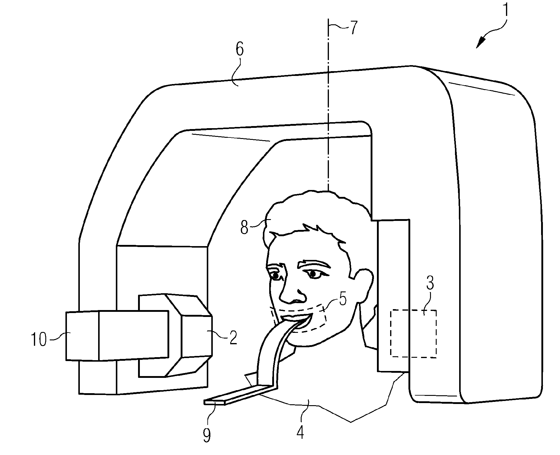
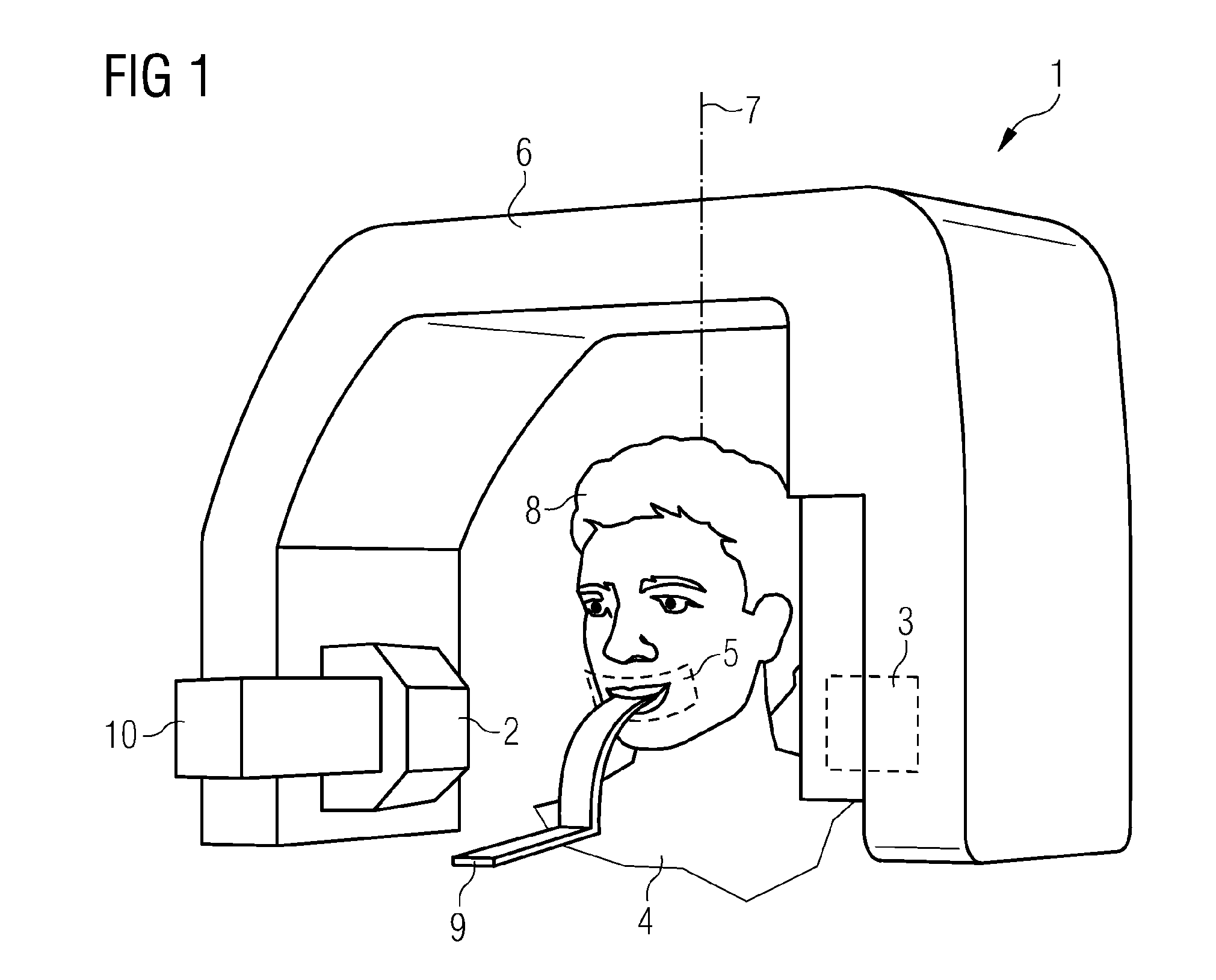
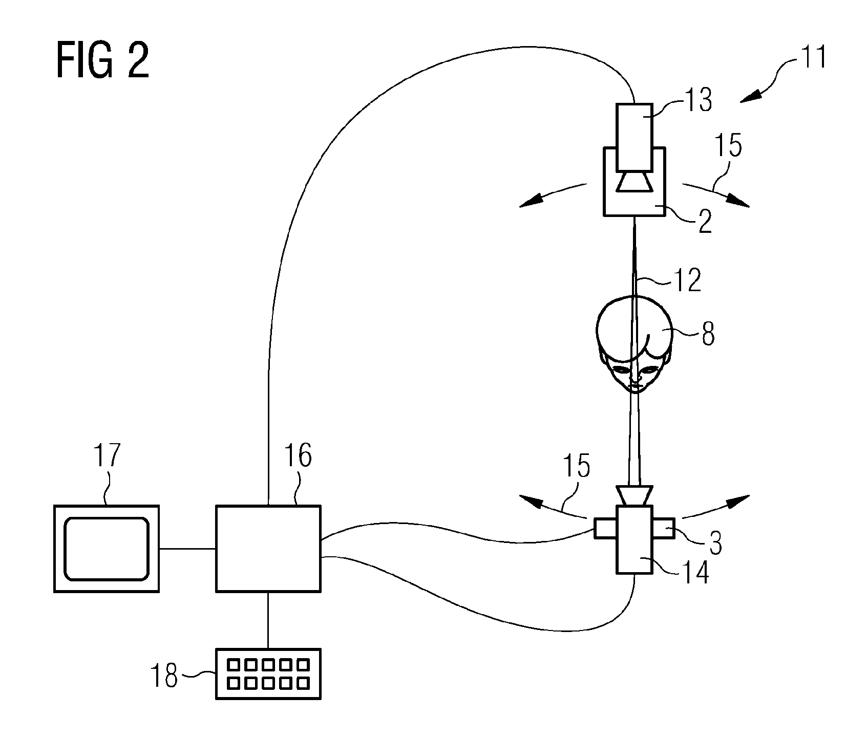
[0060]FIG. 1 shows a apparatus 1 for panoramic dental radiography that comprises a X-ray tube 2 and an X-ray detector 3 for detecting the radiation crossing the body of the patient 4. The detector 3 may be a digital X-ray detector 3 that comprises a matrix of sensible elements or so called pixels, often covered by X-ray detecting scintillator material. Linear shaped area detectors provide the most economic solution to realize a digital radiographic apparatus. For instance, they are widely used to build orthopantomographs aimed to dental radiography. In this case, both the detector 3 and the tube 2 perform a trajectory in the three-dimensional space focusing on a specific target surface 5, for instance the dental arch of the patient 4. For performing the trajectory, the tube 2 and the detector 3 are attached to a support structure 6 that can be rotated around a vertical rotation axis 7 aligned onto a head 8 of the patient 4. For keeping the head 8 of the patient 4, in particular the ...


PUM

PropertyMeasurementUnit
energyaaaaaaaaaa
optical frequencyaaaaaaaaaa
accelerationaaaaaaaaaa

Abstract

A radiographic X-ray apparatus is equipped with multiple devices for the acquisition of anatomical data, in particular cameras. These devices are used to facilitate and automate the imaging process, providing: before exposure the automated identification of the specific anatomical features of the patient and the optimized presetting of the exposure technique factors and projection geometry, tailored on the actual anatomy of the patient; during exposure the optimized X-ray dose modulation, either automatically or selected by the operator, in order to correctly expose the various regions of interest, and accordingly impart reduced dose to other body parts, according to the actual anatomy of the patient and imaging requirements; after exposure the possibility to complement the radiographic image with additional information about the internal and external anatomy, providing valuable tools for the medical analysis and diagnosis.

Description

CROSS-REFERENCE TO RELATED APPLICATIONS[0001]This application claims priority of European Application No. 08 157 813.0 filed on Jun. 6, 2008. The disclosure of this application is hereby incorporated by reference in its entirety, as part of the present disclosure.FIELD OF THE INVENTION[0002]The invention relates to a method for radiographic imaging by means of high energy radiation comprising the method steps of:[0003]positioning a patient by means of a positioning system adapted for positioning a patient with respect to an imaging system comprising a high energy radiation source and a high energy radiation detector;[0004]acquiring multiple biometric data about the actual anatomy of the patient using at least one data acquisition device;[0005]using the imaging system for acquiring radiographic images by an imaging process and setting at least one operational parameter of the imaging process in dependency of the multiple biometric data.BACKGROUND OF THE INVENTION[0006]The invention f...

Claims


Application Information

Patent Timeline
Patent Type & Authority Applications(United States)
IPC IPC(8): A61B6/04G01N23/04A61B6/14G06V10/141G06V10/25
CPCA61B5/0064G06K9/3233A61B5/1072A61B5/1075A61B5/1077A61B5/1176A61B5/4547A61B6/14A61B6/469A61B6/501A61B6/502A61B6/544G06K9/00221G06K9/2027A61B5/107G06V40/16G06V10/141G06V10/25
Inventor BORGHESE, NUNZIO ALBERTOFROSIO, IURINANNI, EROSRINALDI, GERARDOROTONDO, GIUSEPPE
Owner CEFLA SOC COOP
Why Eureka
Try Eureka