Methods of regulating differentiation in mammals

Active Publication Date: 2009-12-17
KOREA ADVANCED INST OF SCI & TECH
0 Cites 12 Cited by

AI-Extracted Technical Summary

Problems solved by technology

In flies, mutations of these factors lead to increased cell proliferation and decreased cell death.
Of the Hippo pathway proteins, only LATS1-, LATS2-, NF2-, and YAP-null mice have been generated; however, these mice are either...

Benefits of technology

[0011]Another embodiment provides a differentiation regulating agent in mammal including one or more selected from the group consisting of a W...

Abstract

Mechanisms regulating cell proliferation stop and differentiation initiation during the development stage of mammalian embryo, and the proteins involved therein, are presented. Differentiation regulators, methods of regulating differentiation, transgenic organisms with loss of expression of the differentiation regulator, and methods of preparing the transgenic organisms, are provided.

Application Domain

Organic active ingredientsPeptide/protein ingredients +13

Technology Topic

OrganismEmbryo +2

Image

  • Methods of regulating differentiation in mammals
  • Methods of regulating differentiation in mammals
  • Methods of regulating differentiation in mammals

Examples

  • Experimental program(16)

Example

Example 1
Generation of WW45 Null Mice
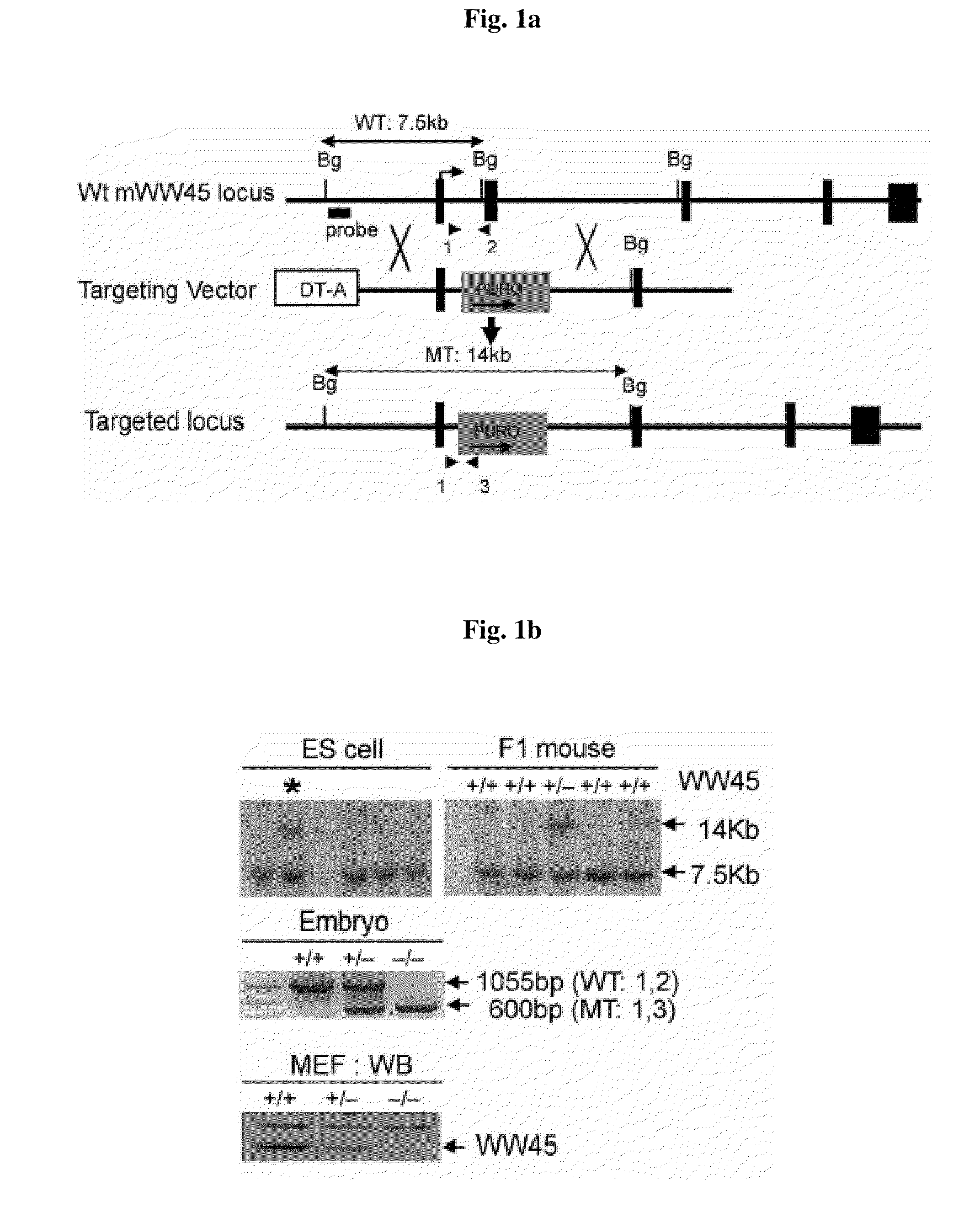
[0115]WW45 gene targeting vector was injected into ES cells by electroporation, and WW45 gene-deficient ES clones were examined by Southern blotting. The WW45 gene-deficient targeting ES clones were injected into blastocyst, to prepare a chimeric mouse.
[0116]More specifically, a WW45−/− mouse was generated using standard ES cell (R1) homologous recombination and blastocyst injection techniques (Lim et al., 1996; Lee et al., 2006). A WW45 targeting vector was prepared by subcloning left arm (obtained from 6338 bp containing exon1 using EcoR I restriction enzyme (New England Biolabs, NEB)) and right arm (obtained from 3443 bp using BamH I restriction enzyme (NEB)) into pGK-Puro vector (obtained from pBluscript II KS(+) vector (Stratagene) as a backbone) using 129SvJ mouse BAC clone (obtained from Korea Research Institute of Bioscience & Biotechnology) (FIG. 1a). More specifically, puromycin resistance gene, which is a positive selection marker, was cloned between left arm and right arm, to replace exon2, and diphtheria toxin-A (DT-A), which is a negative selection marker, was cloned into Not I site, to produce a pGK-Puro-WW45 gene targeting vector.
[0117]The produced WW45 gene targeting vector was deposited with the Korean Collection for Type Cultures (KCTC) at Yuseong-gu, Deajeon, Korea, on Jun. 12, 2008, and allotted receipt number KCTC11342BP.
[0118]WW45−/− mice were generated by injecting the WW45 gene targeting vector into a mouse ES cell (BayGenomics) through electroporation to a targeted ES cell clone, and injecting the ES cell clone into C57BL/6 blastocysts (E3.5, purchased from Jackson Lab) to generate germ-line-transmitting chimeric mice. Mice and ES cells were genotyped by polymerase chain reaction (PCR) assays. The primers and conditions used in the PCR are as follows:
(SEQ ID NO: 13) WW45-L: 5′-TGACCATGTGTCCAGCCTTA-3′, (SEQ ID NO: 14) WW45-R: 5′-CGAATGGATGCTGCATATTG-3′, (SEQ ID NO: 15) pGK-3: 5′-GCACGAGACTAGTGAGACGTGCTAC-3′.
[0119]Conditions: denature at 94° C. for 30 seconds; annealing at 60° C. for 45 seconds; elongation at 72° C. for 1 minute, 30 cycle.
[0120]Using Applied biosystems Geneamp PCR system 2700 PCR apparatus.

Example

Example 2
Histological Analysis
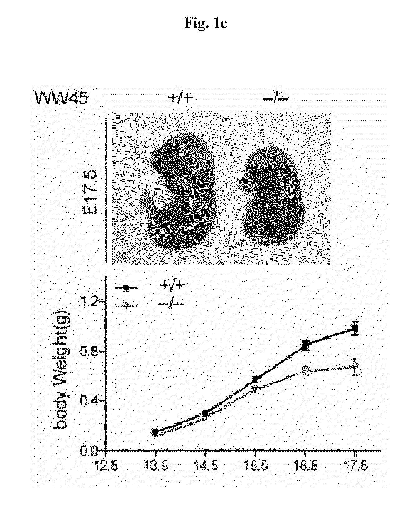
[0121]The chimeric mouse produced in Example 1 was mated with a normal mouse (purchased from C57BL/06, Jackson Lab) to produce WW45 hetero mouse where only one copy of WW45 gene is deleted. It was confirmed by PCR that the embryo obtained by mating the resulted WW45 hetero mice is mutants where the WW45 gene is deleted. The resulted embryo of the transgenic animal was deposited to the Korean Collection for Type Cultures (KCTC) at Yuseong-gu, Deajeon, Korea, on Jun. 12, 2008 and allotted receipt number KCTC11343BP.
[0122]The embryos were fixed in 4% paraformaldehyde overnight at 4° C., embedded in paraffin and sectioned to the size of 4 μm. The sections (4 μm) were stained with hematoxylin and eosin (H&E) or subjected to immunohistochemical analysis. Immunohistochemical analysis was performed using standard protocols (Koo et al., 2007) with antibodies against PECAM-1(BD), laminin (Abcam), BrdU (Sigma), E-cadherin (BD), β-catenin (BD), Ki67 (Novocastra), anti-filaggrin (Covance), loricrin (Covance), K10 (Covance), K14 (Covance), K1 (Covance), iFABP (gift from Dr J. I. Gordon), chromogranin A (Immunostar), YAP (Cell Signaling, Santa Cruz), and p-YAP (Cell Signaling). Peroxidase levels were assessed using the EnVision®+ Dual Link System-HRP(DAB+) (DakoCytomation). Intestinal goblet cells were stained with Alcian blue to detect mucin. The TUNEL assay (koo et al., 2005) was performed using a commercial staining kit (Roche). BrdU incorporation experiments were performed by injecting pregnant females intraperitoneally with BrdU (Sigma) at a concentration of 100 μg/g body weight. At 1 hour, 2 hours, or 24 hours after injection, embryos were dissected and fixed for immunohistochemical analysis.

Example

Example 3
Barrier Function Assays
[0123]At E17.5, the embryos were rinsed in phosphate-buffered saline (PBS) and immersed in acidic X-gal mix (100 mM phosphate buffer at pH4.3, 3 mM K3Fe(CN)6, 3 mM K4Fe(CN)6, 2 mM MgCl2, 1 mg/mL X-gal), then incubated for 8 hours at 37° C. in the dark (Hardman et al., 1998).

PUM

PropertyMeasurementUnit
Length0.0032258m
Fraction1.68fraction
Volume

Description & Claims & Application Information

We can also present the details of the Description, Claims and Application information to help users get a comprehensive understanding of the technical details of the patent, such as background art, summary of invention, brief description of drawings, description of embodiments, and other original content. On the other hand, users can also determine the specific scope of protection of the technology through the list of claims; as well as understand the changes in the life cycle of the technology with the presentation of the patent timeline. Login to view more.
Why Eureka
Try Eureka