

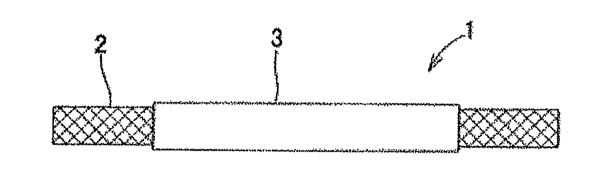
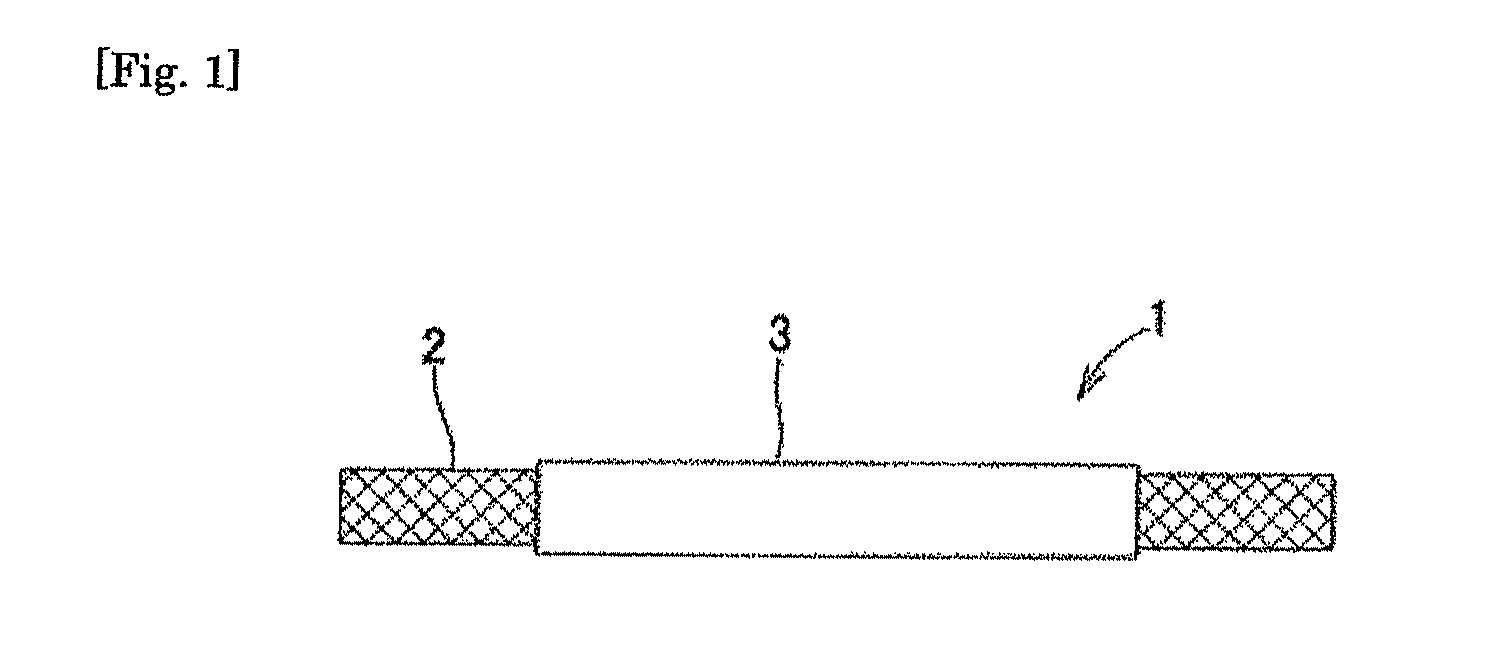
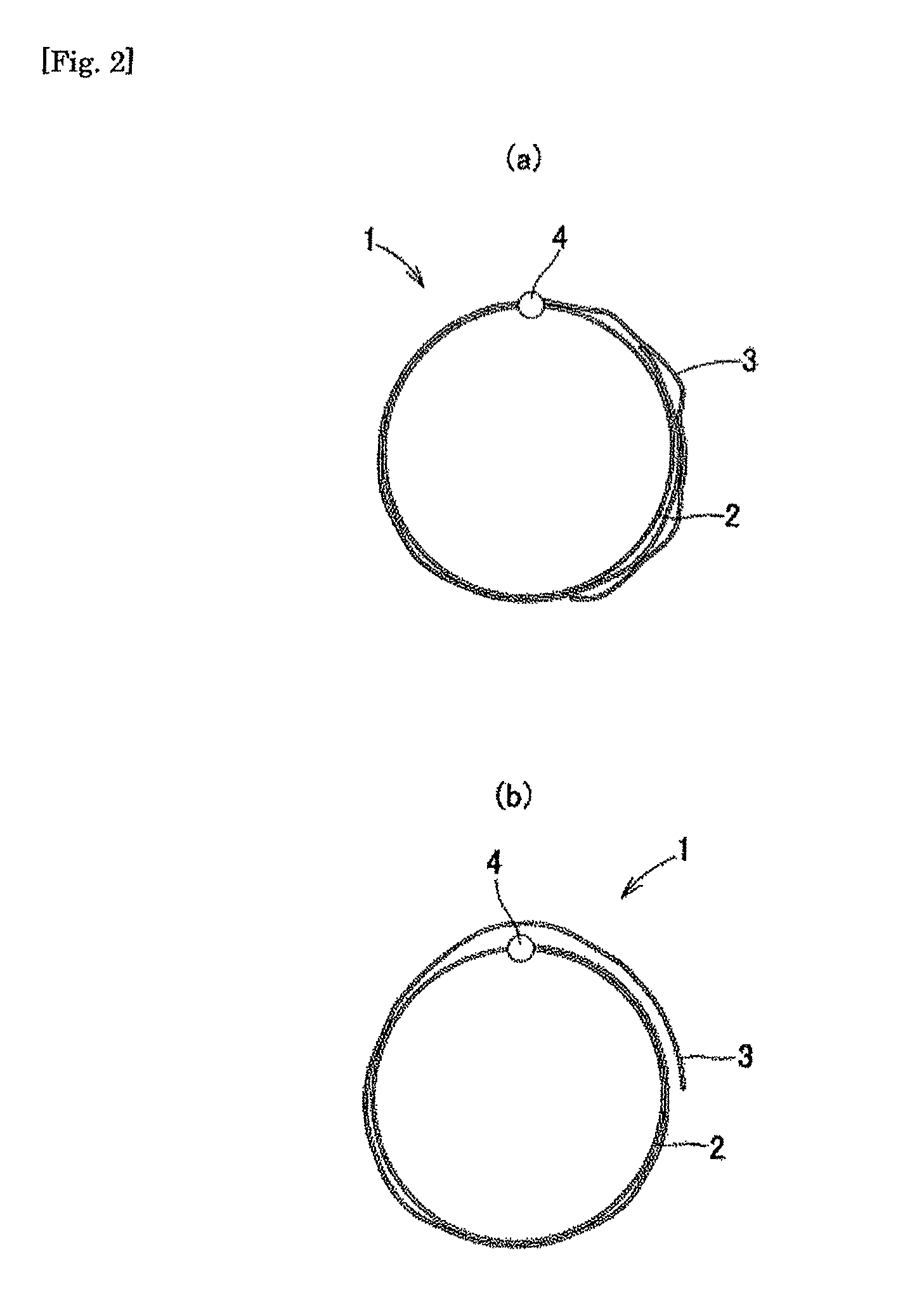
Medical device for body cavity and method of producing the same
a medical device and body cavity technology, applied in the field of new medical devices, can solve the problems of limited blood flow into the aneurysm, unsatisfactory operation convenience, and limited treatment options of aneurysms, so as to reduce the thrombotic debris, and prevent damage to the fragile affected blood vessels
- Summary
- Abstract
- Description
- Claims
- Application Information
AI Technical Summary
Benefits of technology
Problems solved by technology
Method used
Image

Examples
example 1
[0352]FIG. 20 is a slide of the pathological sample of a dog (beagle), after indwelling a medical device, having a plain-woven metal mesh (17×7 mm square, wire diameter: 0.04 mm, thickness: 0.08 mm, size of pore: 0.092 mm, pore rate: 50%, metal (SUS) content: 16.3 mg / mm2) fixed on the peripheral surface of a main stent body (length: 18 mm, balloon expansion diameter: approximately 3.5 mm, made of SUS), to an aneurysm in carotid artery synthetically formed, as shown in FIG. 6. FIG. 20(a) shows a slide of the entire cross section of the blood vessel where the sample stent is indwelled, while FIG. 20(b) is a locally expanded photograph of the slide in FIG. 20(a) (×8 magnification from FIG. 20(a)). It was found at one month after indwelling the medical device that conversion to endodermis was accelerated and the metal mesh 3 and the main stent body 2 were covered with endodermis entirely. FIG. 20(a) was obtained at an objective magnification of ×1.25, and figure (b), at an objective mag...
example 2
[0353]FIG. 21 is photographs of a sample stent after a medical device for body cavity is placed in an aneurysmal phantom and a porcine blood is perfused. In this experiment, a medical device having a plain-woven metal mesh (17×7 mm square, wire diameter: 0.03 mm, thickness 0.05 mm, size of pore: 0.052 mm, pore rate: 43%, metal (SUS) content: 15.5 mg / mm2) fixed on the peripheral surface of a main stent body (length: 18 mm, expansion diameter: approximately 3.5 mm, made of SUS) was indwelled in the aneurysmal phantom, as shown in FIG. 6, and connected to porcine carotid artery for perfusion of blood for 20 minutes. The aneurysmal phantom used was a semi-spherical silicone synthetic aneurysm formed, as adhered to a blood vessel-modeled silicone tube. FIG. 21(a) is a slide of the metal mesh placed in the central region of the aneurysmal opening after perfusion test, and FIG. 21(b) is a SEM photograph of the opening peripheral area of the same synthetic aneurysm in FIG. 21(a). There was ...
example 3
[0354]FIG. 22 is a photograph of a sample stent after a medical device for body cavity is placed in an aneurysmal phantom and a porcine blood is perfused. In this experiment, a medical device having a twill-woven metal mesh (17×7 mm square, wire diameter: 0.02 mm, thickness 0.05 mm, size of pore: 0.03 mm, pore rate: 25%, metal (SUS) content: 11.1 mg / mm2) fixed on the peripheral surface of a main stent body (length: 18 mm, expansion diameter: approximately 3.5 mm, made of SUS) was indwelled in the aneurysmal phantom, as shown in FIG. 6, and connected to porcine carotid artery for perfusion of blood for 20 minutes. The aneurysmal phantom used was a semi-spherical silicone synthetic aneurysm formed, as adhered to a blood vessel-modeled silicone tube.
[0355]FIG. 22(a) is a photograph of the opening-sided aneurysmal phantom after perfusion experiment. FIG. 22(a) showed a great amount of thrombus deposited.
[0356]FIG. 22(b) is a SEM photograph of the artery phantom-sided surface of the mesh...
PUM
 Abstract
Description
Claims
Application Information