Detecting optical properties of a turbid medium

a technology of optical properties and turbid mediums, applied in the field of detecting optical properties of turbid mediums, to achieve the effects of improving the correlation between optical properties and disease state, improving diagnostic accuracy, and improving diagnostic accuracy

Active Publication Date: 2010-02-18
TRUSTEES OF BOSTON UNIV
View PDF5 Cites 21 Cited by
  • Summary
  • Abstract
  • Description
  • Claims
  • Application Information

AI Technical Summary

Benefits of technology

[0029]In another embodiment of this and all other aspects described herein, the turbid medium of interest and the reference turbid medium are each an epithelial layer, and a difference in the absolute value is indicative of a physiological difference between the epithelial layer in the sample of interest and the reference. The physiological difference can be indicative of a disease state. The methods and devices described herein are well suited for the situation in which a test determination of optical properties of a tissue provide absolute values that can be compared to standards to provide a direct diagnostic tool, i.e., an indicator of disease. The indicator can either point directly to a disease state—i.e., no further testing is required, or, alternatively, can provide information suggestive of a disease state, i.e., an indication that further, potentially more invasive testing is warranted, such as a biopsy. Better correlations between differences in optical properties and disease state can be gained where a larger number of samples are examined to establish both healthy and disease tissue (e.g, cancer, irritable bowel syndrome, inflammatory disease, Crohn's disease, cervical disease, etc.) standards. Many diseases are correlated with the optical properties (scattering and absorption) of tissue. The scattering is dependent on the morphological composition of cell scattering centers, such as nucleus, mitochondria and other organelles. The absorption is dependent on the concentration of several endogenous compounds such as hemoglobin, bilirubin and cytochrome oxidase, and exogenous compounds such as several chemotherapy and photodynamic therapy agents. Most cancers originate in the epithelium (a superficial tissue layer with a thickness of 100-500 μm) of organs such as the esophagus, skin and urinary bladder. A standard or set of standards based on a large sample size can thus provide a more reliable diagnostic criterion for comparison. When a test sample varies from a normal sample in a degree to which a known disease sample or standard does, the test can correlate very accurately with disease diagnosis. Thus the degree of variation that is considered a “difference” will generally depend upon the sample size of the reference or standard, with larger sample numbers for both standard tissues (i.e., known healthy or known diseased) and test tissues providing more sensitive results.

Problems solved by technology

Whereas relative values are of somewhat limited use in that they do not readily translate to comparisons with other samples, an absolute value can be compared effectively with an absolute value from another sample with meaningful results.

Method used


Image

  • Detecting optical properties of a turbid medium
  • Detecting optical properties of a turbid medium
  • Detecting optical properties of a turbid medium

Examples


example 1a

Exemplary Methods for Defining an Analytical Model Described Herein

A. Monte Carlo Simulations

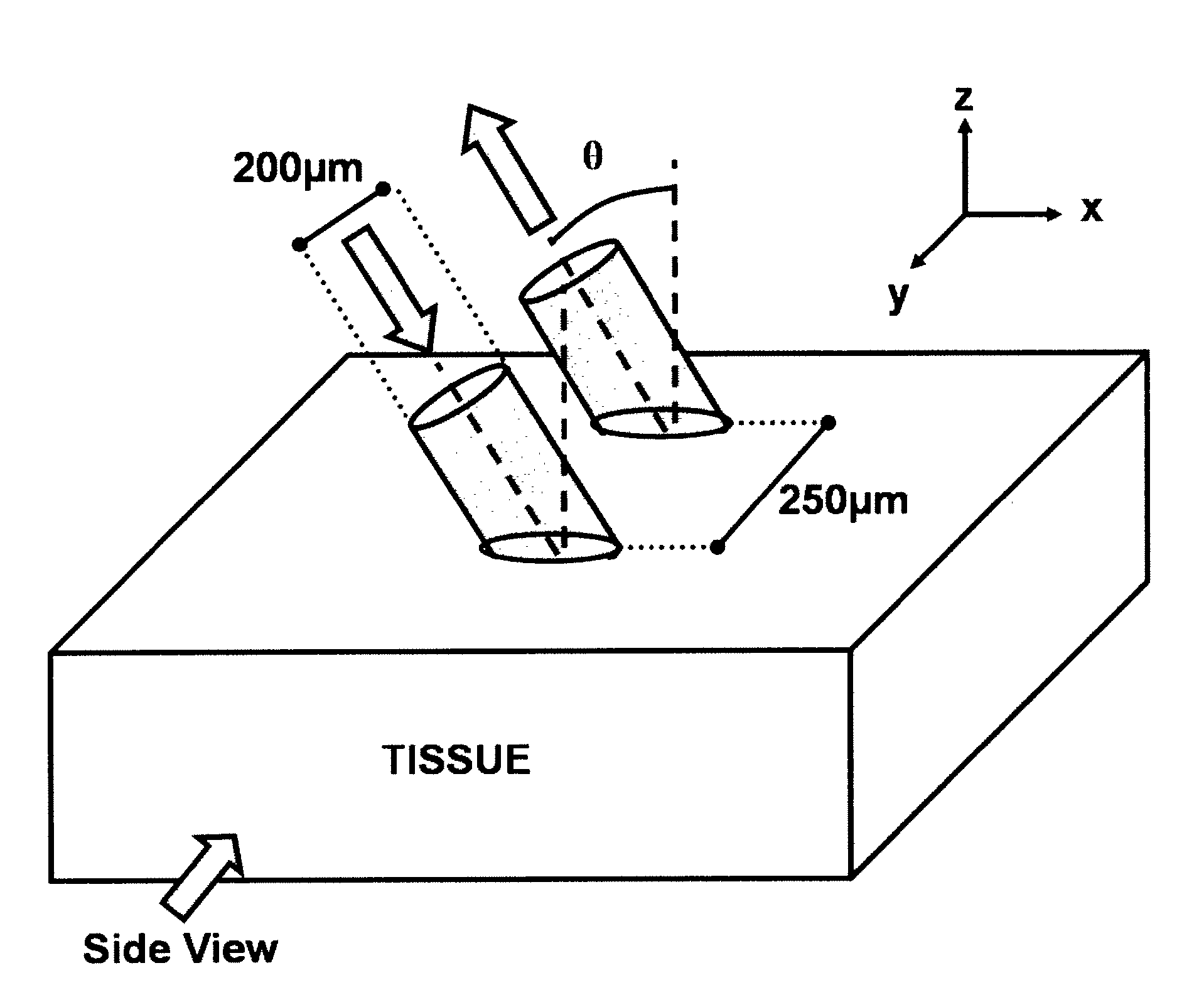
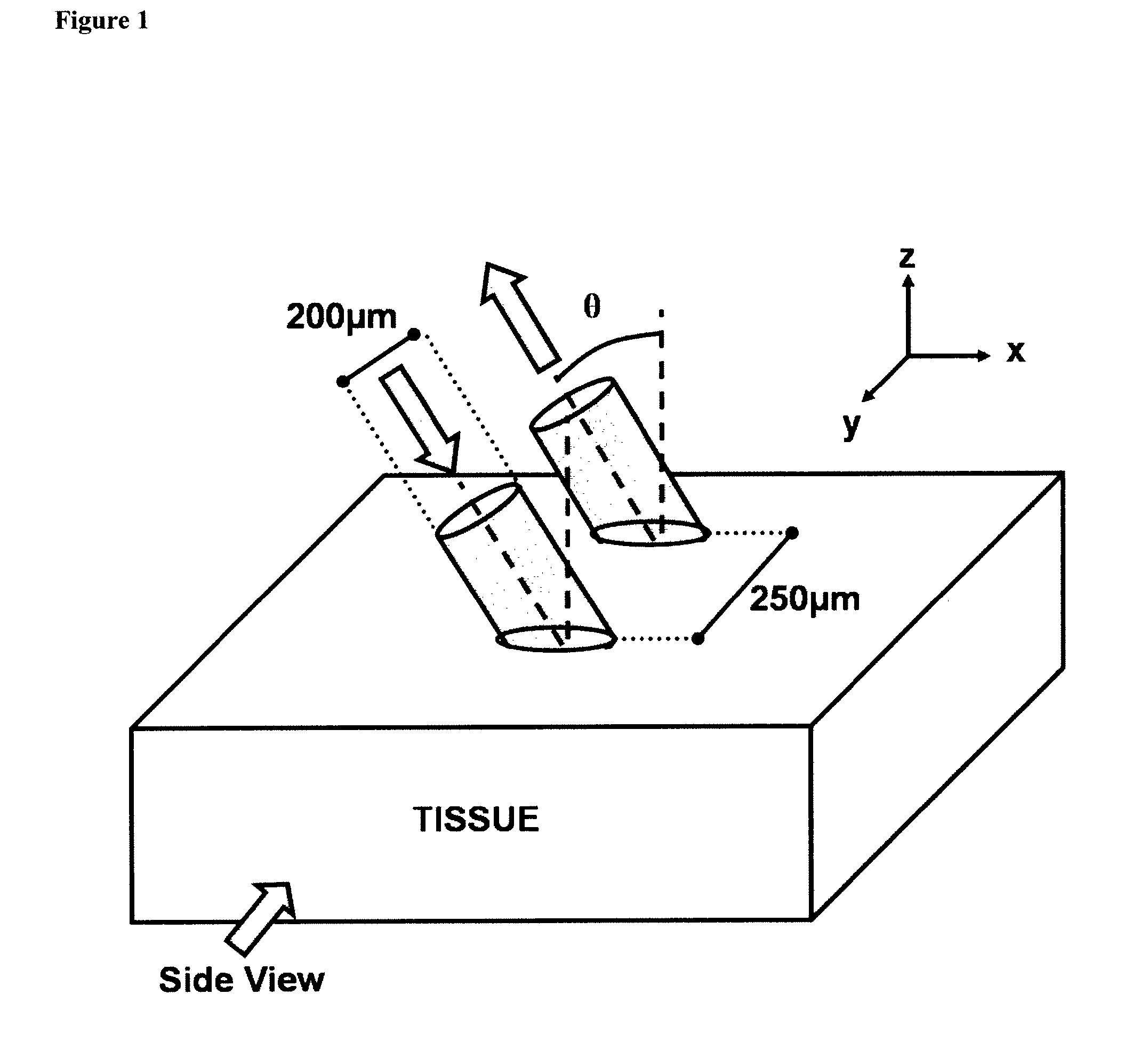
[0060]A Monte Carlo (MC) code that simulates light transport within a scattering and absorbing medium has been developed based on previous codes31,32, using a variance reduction technique33. The MC code simulates a probe that comprises two fibers (a source and a detector), as depicted in FIG. 1. Each fiber has a core diameter of 200 μm and a numerical aperture (NA) of 0.22 in air. The fibers have a center-to-center separation of 250 μm. Both fibers can be tilted at an angle (θ) relative to the tissue surface normal, in the x-z plane, such that the fibers are parallel to each other. Four tilt angles have been investigated: 0, 15, 30 and 45 degrees. The surfaces of the fibers were modeled to be polished at an angle such that the faces of the fibers were parallel to the surface of the medium. Thus, the faces of the fibers had a circular or elliptical shape depending on whether θ=0 or θ>0, respe...

example 1b

Development of an Analytical Model as Described Herein

A. Reflectance as a Function of the Reduced Scattering Coefficient

[0070]For the development of the model described herein, the reflectance from a medium that scatters light was analyzed for conditions with very little absorption (μa1≦0.01 cm−1). MC simulations were run for a medium with an absorption coefficient of 0.01 cm−1 and for five values of reduced scattering coefficient (μs′=5, 10, 15, 20 and 25 cm−1). The anisotropy value was set to 0.9. Four different fiber tilt angles were modeled (θ=0, 15, 30 and 45 degrees). The absolute reflectance (RABS), is defined as the ratio of the collected light (I) over the incident light (I0), and is calculated using Eq. (1):

RABS=II0=∑i=1TPCexp(-μali)TPL(1)

where TPC is the Total number of Photons Collected, TPL is the Total number of Photons Launched, μa is the absorption coefficient and li is the path-length of each collected photon. To analyze the reflectance experimentally, eight tissue ...

example 2

An Exemplary Model for Extracting Absolute Optical Properties of a Biological Tissue

[0111]Non-invasive optical reflectance measurements have been used to extract diagnostic information from a variety of tissue types, in vivo, such as colon49, skin50, brain51, cervix52, esophagus53, prostate54 and bronchial mucosa55. The typical experimental setup of these systems often consists of a fiber probe with a diameter of a few millimeters or less, with one or more fibers that transmit light to and from the tissue. An earlier ex vivo study reported that the amount of pressure applied on a sample of tissue affects its absorption and reduced scattering coefficients56. It has also been reported that probe pressures with forces of less than 1 Newton do not affect the fluorescence intensity or lineshape of measurements obtained in vivo on cervical tissues57.

[0112]Described herein is a model of the influence of probe pressure on spectral reflectance measurements of biological tissue in vivo. The t...


PUM

PropertyMeasurementUnit
diameteraaaaaaaaaa
thicknessaaaaaaaaaa
distancesaaaaaaaaaa

Abstract

A device for measuring light scattering and absorption properties of a tissue, the device comprising: a probe comprising first and second optical fibers for irradiation and detection. The optical fibers are substantially parallel to each other at the distal end, and are separated by a distance of less than 2 mm, wherein said first and second optical fibers are arranged in said probe at an angle, 6=10 deg. to 45 deg., to the plane perpendicular to the distal end of said probe and wherein the tips of the optical fibers at the distal end are polished parallel to the plane perpendicular to the distal end of said probe.

Description

CROSS-REFERENCE TO RELATED APPLICATIONS[0001]The present application claim benefit under 35 U.S.C. §119(e) of U.S. Provisional Application Ser. No. 60 / 920,410 filed Mar. 28, 2007, the content of which is herein incorporated by reference in its entirety.GOVERNMENT SUPPORT[0002]This invention was made with Government support under grant numbers U54 CA104677-01 and F31 CA110016 awarded by the National Institutes of Health. The Government has certain rights in this invention.FIELD OF THE INVENTION[0003]The present invention relates to detecting optical properties of a turbid medium. The invention also relates to extracting optical properties from data obtained from biological tissue.BACKGROUND[0004]An important challenge in biomedical research is to non-invasively characterize tissue conditions, for the purposes of diagnosis of disease, such as (pre)cancerous conditions, or for monitoring response to treatment. Non-invasive optical reflectance measurements have been used to extract diag...

Claims


Application Information

Patent Timeline
Patent Type & Authority Applications(United States)
IPC IPC(8): A61B6/00G01N21/00G01N21/55
CPCA61B5/0075G01N21/59G01N21/49A61B5/0084
Inventor BIGIO, IRVING J.REIF, ROBERTOA'AMAR, OUSAMA
Owner TRUSTEES OF BOSTON UNIV
Why Eureka
Try Eureka