Detecting optical properties of a turbid medium
a technology of optical properties and turbid mediums, applied in the field of detecting optical properties of turbid mediums, to achieve the effects of improving the correlation between optical properties and disease state, improving diagnostic accuracy, and improving diagnostic accuracy
- Summary
- Abstract
- Description
- Claims
- Application Information
AI Technical Summary
Benefits of technology
Problems solved by technology
Method used
Image
Examples
example 1a
Exemplary Methods for Defining an Analytical Model Described Herein
A. Monte Carlo Simulations
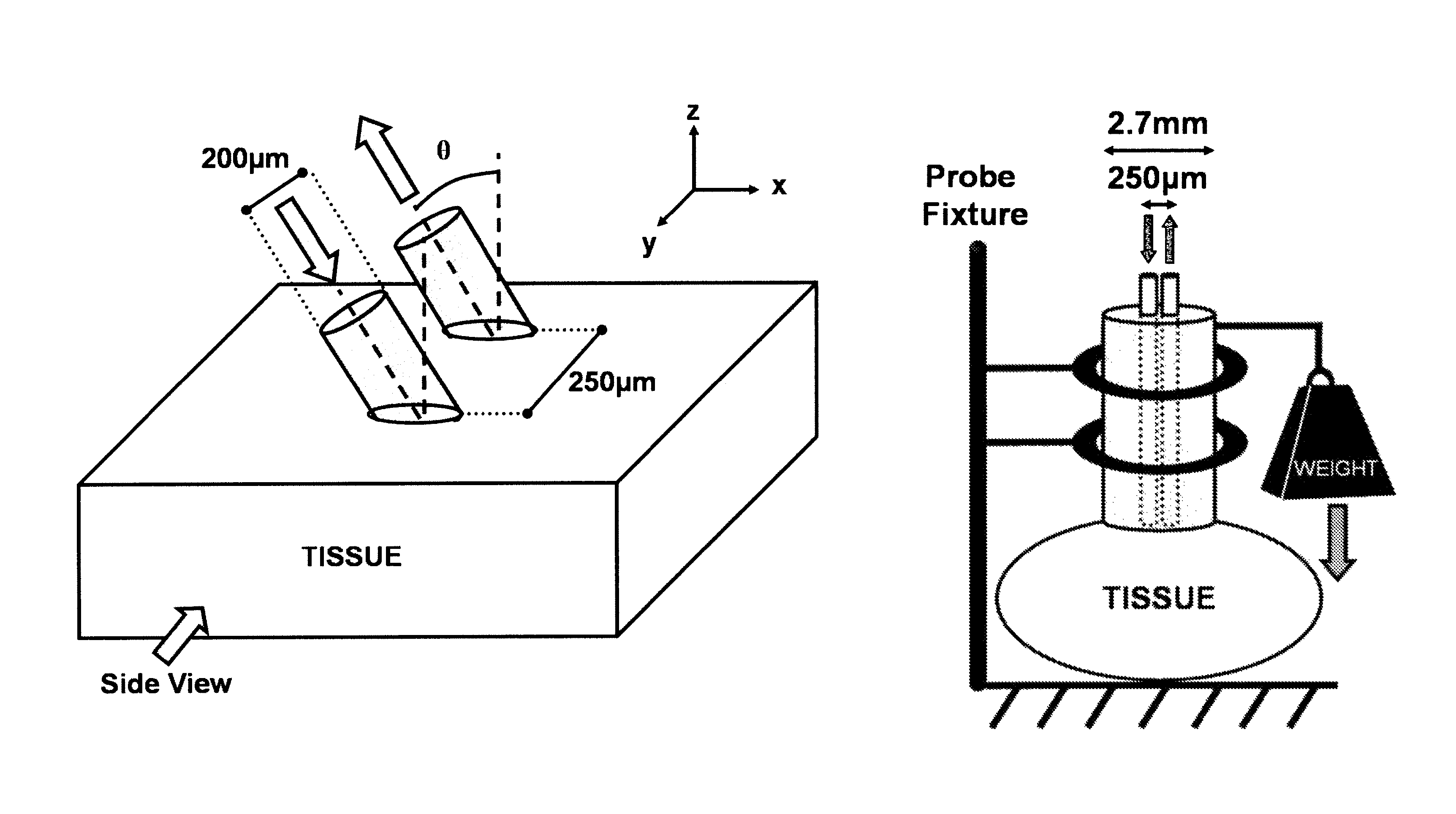
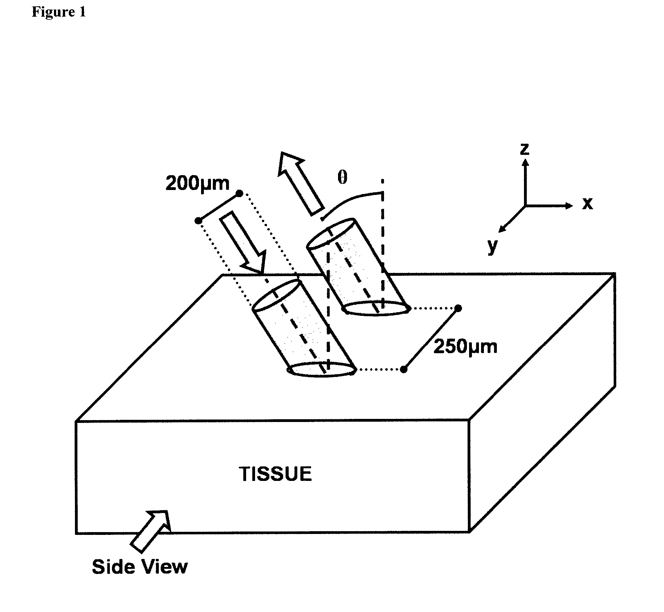
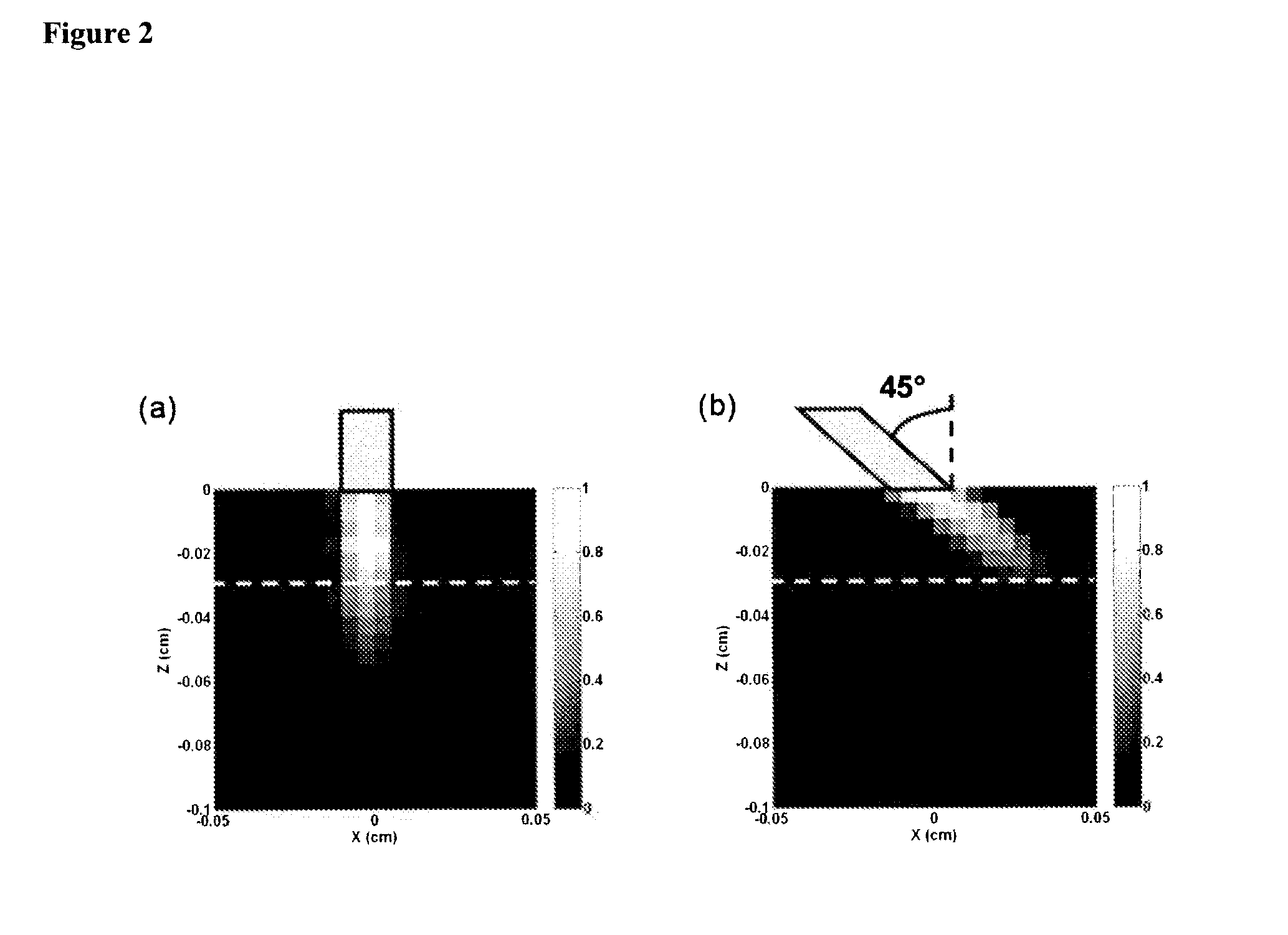
[0072]A Monte Carlo (MC) code that simulates light transport within a scattering and absorbing medium has been developed based on previous codes31,32, using a variance reduction technique33. The MC code simulates a probe that comprises two fibers (a source and a detector), as depicted in FIG. 1. Each fiber has a core diameter of 200 μm and a numerical aperture (NA) of 0.22 in air. The fibers have a center-to-center separation of 250 μm. Both fibers can be tilted at an angle (θ) relative to the tissue surface normal, in the x-z plane, such that the fibers are parallel to each other. Four tilt angles have been investigated: 0, 15, 30 and 45 degrees. The surfaces of the fibers were modeled to be polished at an angle such that the faces of the fibers were parallel to the surface of the medium. Thus, the faces of the fibers had a circular or elliptical shape depending on whether θ=0 or θ>0, respe...
example 1b
Development of an Analytical Model as Described Herein
A. Reflectance as a Function of the Reduced Scattering Coefficient
[0082]For the development of the model described herein, the reflectance from a medium that scatters light was analyzed for conditions with very little absorption (μa1≦0.01 cm−1). MC simulations were run for a medium with an absorption coefficient of 0.01 cm−1 and for five values of reduced scattering coefficient (μs′=5, 10, 15, 20 and 25 cm−1). The anisotropy value was set to 0.9. Four different fiber tilt angles were modeled (θ=0, 15, 30 and 45 degrees). The absolute reflectance (RABS), is defined as the ratio of the collected light (I) over the incident light (I0), and is calculated using Eq. (1):
[0083]RABS=II0=∑i=1TPCexp(-μali)TPL(1)
where TPC is the Total number of Photons Collected, TPL is the Total number of Photons Launched, μa is the absorption coefficient and li is the path-length of each collected photon. To analyze the reflectance experimentally, eigh...
example 2
An Exemplary Model for Extracting Absolute Optical Properties of a Biological Tissue
[0134]Non-invasive optical reflectance measurements have been used to extract diagnostic information from a variety of tissue types, in vivo, such as colon49, skin50, brain51, cervix52, esophagus53, prostate54 and bronchial mucosa55. The typical experimental setup of these systems often consists of a fiber probe with a diameter of a few millimeters or less, with one or more fibers that transmit light to and from the tissue. An earlier ex vivo study reported that the amount of pressure applied on a sample of tissue affects its absorption and reduced scattering coefficients56. It has also been reported that probe pressures with forces of less than 1 Newton do not affect the fluorescence intensity or lineshape of measurements obtained in vivo on cervical tissues57.
[0135]Described herein is a model of the influence of probe pressure on spectral reflectance measurements of biological tissue in vivo. The t...
PUM
 Abstract
Description
Claims
Application Information
 - R&D
- Intellectual Property
- Life Sciences
- Materials
- Tech Scout
- Unparalleled Data Quality
- Higher Quality Content
- 60% Fewer Hallucinations
Browse by: Latest US Patents, China's latest patents, Technical Efficacy Thesaurus, Application Domain, Technology Topic, Popular Technical Reports.
© 2025 PatSnap. All rights reserved.Legal|Privacy policy|Modern Slavery Act Transparency Statement|Sitemap|About US| Contact US: help@patsnap.com


