System, Method and Apparatus for Measuring Blood Flow and Blood Volume

Active Publication Date: 2010-03-18
BAXTER INT INC +1
View PDF66 Cites 21 Cited by
  • Summary
  • Abstract
  • Description
  • Claims
  • Application Information

AI Technical Summary

Benefits of technology

[0079]According to still further features in the described preferred embodiments the electronic circuitry comprises a digitizer for digitizing the remaining portion of the mixed radiofrequency signal. According to still further features in th

Problems solved by technology

Generally, heart diseases may be caused by (i) a failure in the autonomic nerve system where the impulses from the central nervous system control to the heart muscle fail to provide a regular heart rate and/or (ii) an insufficient strength of the heart muscle itself where even though the patient has a regular heart rate, its force of contraction is insufficient.
A common clinical problem is that of hypotension (low blood pressure); this may occur because the cardiac output is low and/or because of low systemic vascular resistance.
This problem can occur in a wide range of patients, especially those in intensive care or postoperative high dependency units.
The Fick method suffers from many drawbacks.
First, accurate collection of the gas is difficult unless the patient has an endotracheal tube because of leaks around a facemask or mouthpiece.
Second, the analysis of the gas, which is straightforward if the inspired gas is air, is problematic for oxygen enriched air.
Third, the arteriovenous oxygen content difference presents a further problem in that the mixed venous (i.e., pulmonary arterial) oxygen content has to be measured and therefore a pulmonary artery catheter is needed to obtain the sample, which may cause complications to the patient.
Although this method improves the ability to perform accurate measurements of gas, it still suffers from most of the above limitations, in particular the limitation related to leaks around the facemask.
Besides being very inaccurate, the method has the following disadvantages: (i) the system may only be operated by a skilled operator; (ii) due to the size of the system's probe, heavy sedation or anaesthesia is needed; (iii) the system is expensive; and (iv) the probe cannot be configured to provide continuous cardiac output readings without an expert operator being present.
This method, however, is very inaccurate and it can only provide a rough estimation of the

Method used


Image

  • System, Method and Apparatus for Measuring Blood Flow and Blood Volume
  • System, Method and Apparatus for Measuring Blood Flow and Blood Volume
  • System, Method and Apparatus for Measuring Blood Flow and Blood Volume

Examples


example 1

Measurement of Stroke Volume and Cardiac Output Using Three Electrodes

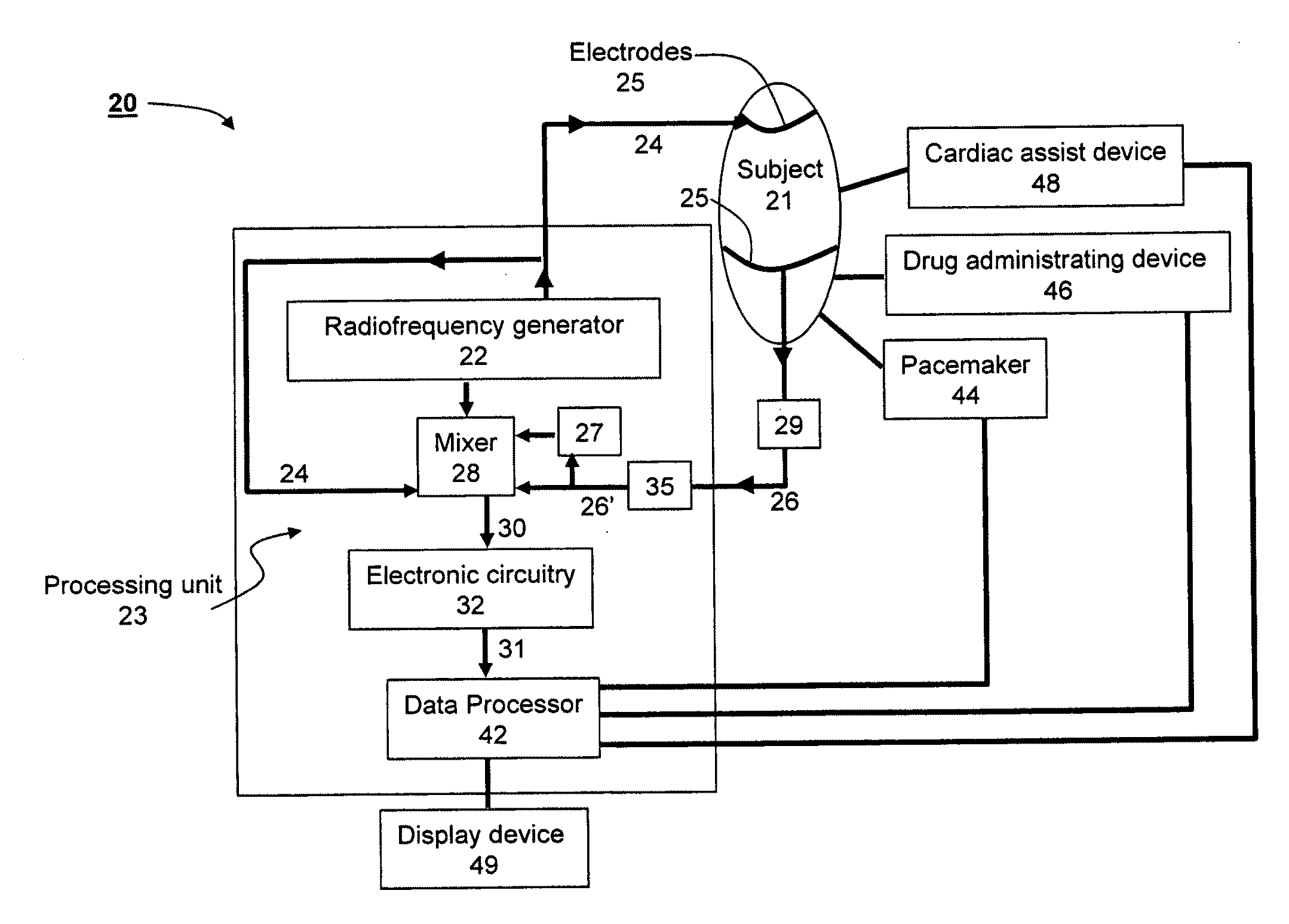
[0185]Three electrodes were connected to a human subject, as shown in FIG. 4a. The hemodynamic reactance was measured and was used for determining and monitoring (i) stroke volume; and (ii) cardiac output.

[0186]FIGS. 10a-b shows the monitoring results obtained using the prototype system (using the circuitry of FIG. 9a) on a time scale of 250 ms / div. Two waveforms are displayed in each of FIGS. 10a-b, the change in the hemodynamic reactance and its measured time derivative. The waveforms shown in FIG. 10b are in reverse magnification compared to the waveforms shown in FIG. 10a.

[0187]For comparison, FIG. 10c shows monitoring results obtained using a conventional system (GE / Cardiodynamic). The waveforms displayed in FIG. 10c, are, from top to bottom, the ECG signal, the change in the bioimpedance, ΔZ, its first derivative, dZ / dt and its second derivative d2Z / dt2.

[0188]The improvement of the signal-to noise ratio of ...

example 2

Measurement of Brain Intra Luminal Blood Volume Change and Flow Rate Using Two Electrodes

[0189]Two electrodes were connected to a human subject, as shown in FIG. 4e. The hemodynamic reactance was measured and was used for determining and monitoring brain intra luminal blood volume change and flow rate.

[0190]FIGS. 11a-b show the monitoring results obtained using the prototype system (using the circuitry of FIG. 9b) on a time scale of 250 ms / div. Two waveforms are displayed in each of FIGS. 11a-b, the change in the hemodynamic reactance and its measured derivative, where in FIG. 11b, the vertical scale for the curve of the change in the hemodynamic reactance is twice larger than the respective curve in

[0191]FIG. 11a.

[0192]As shown in FIGS. 11a-b, a good signal-to noise ratio of 50 dB was obtained for both quantities. The curves of the present example acquire a sharper peak, as compared to Example 1. This phenomenon is consistent with physiological findings, according to which the res...

example 3

Measurement of Stroke Volume and Cardiac Output Using Four Electrodes

[0193]Four electrodes were connected to a human subject, as shown in FIG. 4b. The hemodynamic reactance was measured and was used for determining and monitoring (i) stroke volume; and (ii) cardiac output.

[0194]FIG. 12a shows the monitoring results obtained using the prototype system (using the circuitry of FIG. 9c) on a time scale of 500 ms / div. Two waveforms are displayed in FIG. 12, the change in the change in the hemodynamic reactance and its measured time derivative.

[0195]FIG. 12b shows a comparison between the CO signal as calculated from the phase shift Δφ according to the embodiments of the invention, and data acquired from other channels. From top to bottom, FIG. 12b shows, as a function of time: ECG lead I (designated I In FIG. 12b), ECG lead II (designated II), left blood wave front (L), right blood wave front (R), CO signal (N), first derivative of the CO signal (dN) and second derivative of the CO signa...


PUM


Abstract

A method of calculating blood flow in an organ of a subject using output radiofrequency signals transmitted to the organ and input radiofrequency signals received from the organ, the method comprises determining a phase shift of the input radiofrequency signals relative to the output radiofrequency signals and using the phase shift to calculate the blood flow in the organ.

Description

FIELD AND BACKGROUND OF THE INVENTION[0001]The present invention relates to measurement of electrical signals of a body of a subject and, more particularly, to measurement of electrical signals of the body of the subject so as to determine blood volume or blood volume rate, e.g., stroke volume, cardiac output, brain intra luminal blood volume and the like.[0002]Heart diseases are major causes of morbidity and mortality in the modern world. Generally, heart diseases may be caused by (i) a failure in the autonomic nerve system where the impulses from the central nervous system control to the heart muscle fail to provide a regular heart rate and / or (ii) an insufficient strength of the heart muscle itself where even though the patient has a regular heart rate, its force of contraction is insufficient. Either way, the amount of blood or the rate at which the blood is supplied by a diseased heart is abnormal and it is appreciated that an assessment of the state of a patient's circulation ...

Claims


Application Information

Patent Timeline
IPC IPC(8): A61B5/02
CPCA61B5/0535A61B5/026A61B5/7239A61B5/0295
Inventor KEREN, HANAN
Owner BAXTER INT INC
Why Eureka
Try Eureka