
Method for magnitude constrained phase contrast magnetic resonance imaging

Active Publication Date: 2010-04-08
WISCONSIN ALUMNI RES FOUND
View PDF7 Cites 26 Cited by
  • Summary
  • Abstract
  • Description
  • Claims
  • Application Information

AI Technical Summary

Benefits of technology

[0019]It is an aspect of the present invention to provide a method for reconstructing high resolution angiographic velocity images that employs non-Cartesian data acquisition schemes. Such a method creates a tradeoff between image artifacts from off-resonance effects and aliasing artifacts from fat. Particularly in body imaging, fat content can cause degraded image quality, create errors in the velocity measurements, and prevent the use of self-calibrated B0 inhomogeneity field maps. The influence of fat is reduced and self-calibrated B0 inhomogeneity corrections are facilitated by employing a combination of chemical shift imaging with phase contrast velocimetry with non-linear least squares estimation of velocity, fat, and water signals.

Problems solved by technology

Such a method creates a tradeoff between image artifacts from off-resonance effects and aliasing artifacts from fat.
Particularly in body imaging, fat content can cause degraded image quality, create errors in the velocity measurements, and prevent the use of self-calibrated B0 inhomogeneity field maps.

Method used


Image

  • Method for magnitude constrained phase contrast magnetic resonance imaging
  • Method for magnitude constrained phase contrast magnetic resonance imaging
  • Method for magnitude constrained phase contrast magnetic resonance imaging

Examples


Embodiment Construction

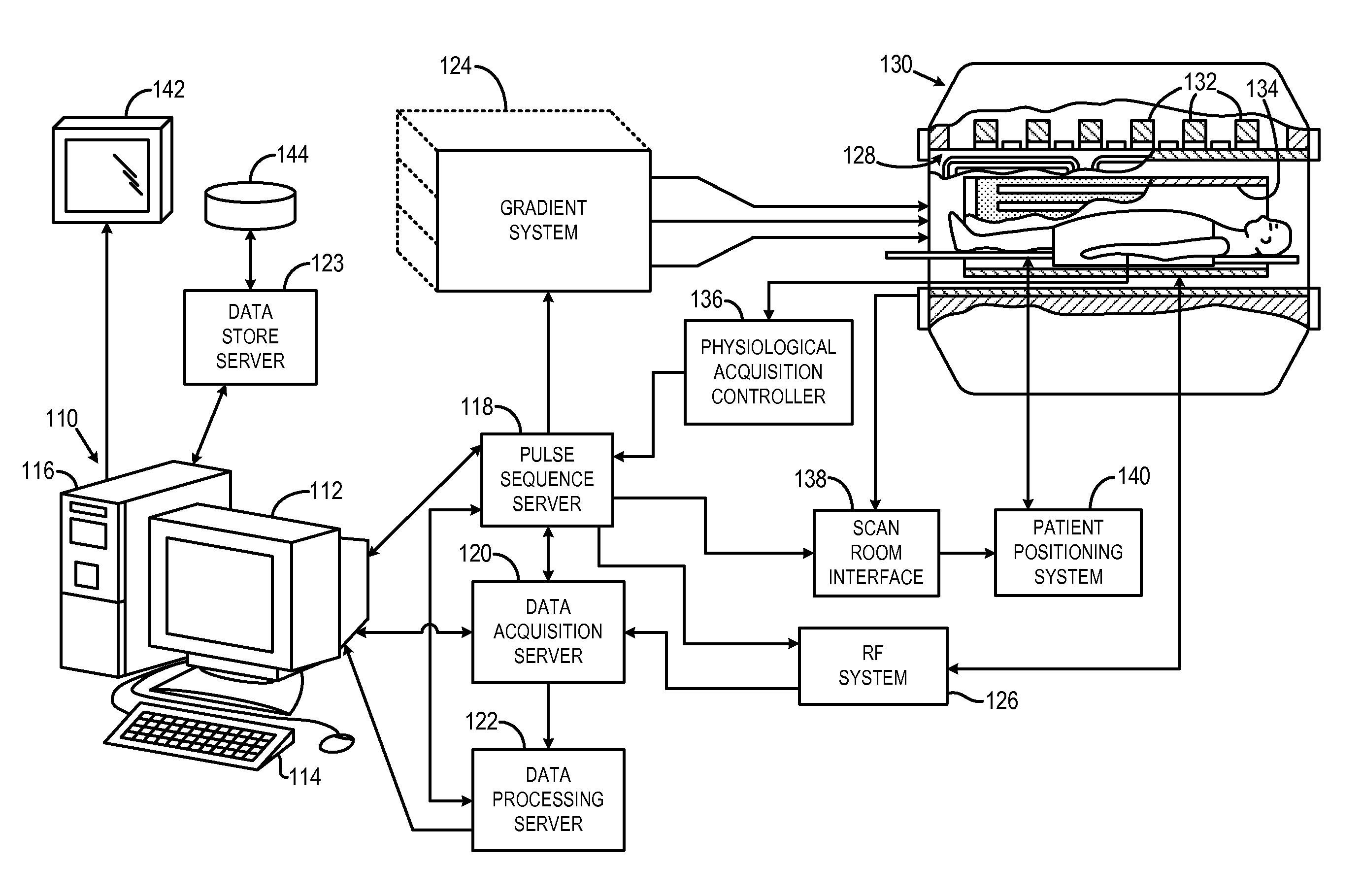
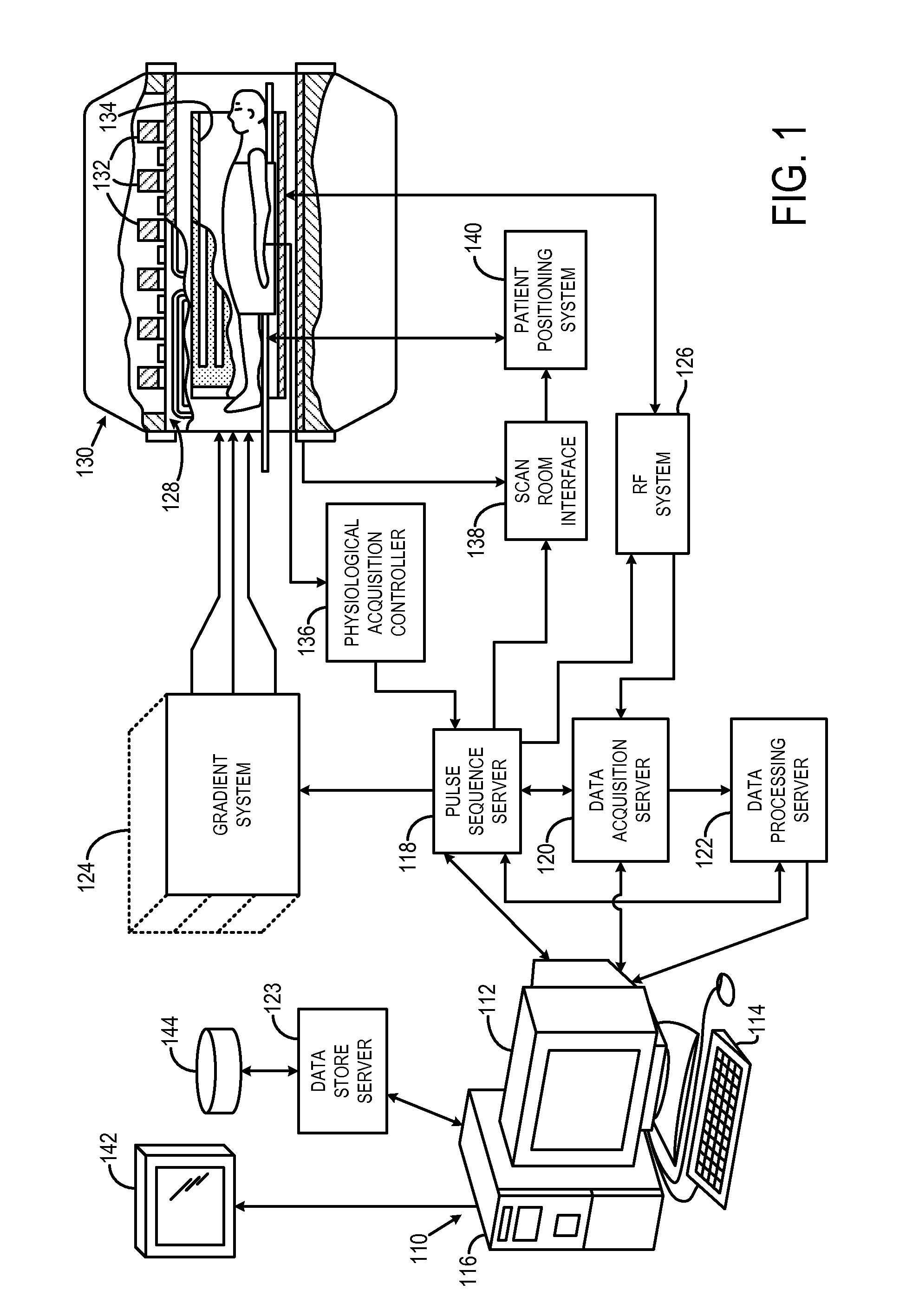
[0027]Referring particularly to FIG. 1, the preferred embodiment of the invention is employed in a magnetic resonance imaging (“MRI”) system. The MRI system includes a workstation 110 having a display 112 and a keyboard 114. The workstation 110 includes a processor 116 that is a commercially available programmable machine running a commercially available operating system. The workstation 110 provides the operator interface that enables scan prescriptions to be entered into the MRI system. The workstation 110 is coupled to four servers: a pulse sequence server 118; a data acquisition server 120; a data processing server 122, and a data store server 123. The workstation 110 and each server 118, 120, 122 and 123 are connected to communicate with each other.

[0028]The pulse sequence server 118 functions in response to instructions downloaded from the workstation 110 to operate a gradient system 124 and a radio frequency (“RF”) system 126. Gradient waveforms necessary to perform the presc...


PUM


Abstract

A method for magnitude constrained phase contrast magnetic resonance imaging (MRI) is provided. The method utilizes an assumption that the image magnitude is shared across a series of images reconstructed from a set of phase contrast enhanced k-space data. In this manner, one common magnitude image and a plurality of phase images are reconstructed substantially contemporaneously from the acquired image data. The method is further applicable to other phase contrast MRI methods, such as phase contract velocimetry. Moreover, simultaneous phase contrast velocimetry and chemical shift imaging, in which water and fat signal separation is achieved, is provided.

Description

CROSS-REFERENCE TO RELATED APPLICATIONS[0001]This application claims the benefit of U.S. Provisional Patent Application Ser. No. 61 / 102,712 filed on Oct. 3, 2008, and entitled “Method for Magnitude Constrained Phase Contrast Magnetic Resonance Imaging.”STATEMENT REGARDING FEDERALLY SPONSORED RESEARCH[0002]This invention was made with United States government support awarded by the following agency: NIH CA116380. The United States government has certain rights in this invention.BACKGROUND OF THE INVENTION[0003]The field of the invention is magnetic resonance imaging (“MRI”) methods and systems. More particularly, the invention relates to methods and systems for phase contrast MRI.[0004]When a substance such as human tissue is subjected to a uniform magnetic field (polarizing field B0), the individual magnetic moments of the nuclei in the tissue attempt to align with this polarizing field, but precess about it in random order at their characteristic Larmor frequency. If the substance,...

Claims


Application Information

Patent Timeline
IPC IPC(8): G01R33/48
CPCG01R33/4824G01R33/4828G01R33/56563G01R33/56308G01R33/56316G01R33/561
Inventor JOHNSON, KEVIN M.SAMSONOV, ALEXEY A.
Owner WISCONSIN ALUMNI RES FOUND
Learn More