Methods for reducing the side effects of ophthalmic laser surgery

a laser surgery and laser technology, applied in the field of stem cell therapy, can solve the problems of retinal detachment, vitreous hemorrhage, and retinal neovascularization often causing eye bleeding, and achieve the effects of reducing the risk of ophthalmic laser surgery side effects, and reducing the risk of ophthalmic surgery side effects

Inactive Publication Date: 2010-04-22
TANKOVICH NIKOLAI
View PDF3 Cites 10 Cited by
  • Summary
  • Abstract
  • Description
  • Claims
  • Application Information

AI Technical Summary

Benefits of technology

[0010]One objective of the invention is to provide a method for treating a side effect resulting from the application of laser therapy in the treatment of an eye disorder comprising treating an eye disorder in a patient with a laser, wherein said treating produces a side effect in the eye of said patient, administering a therapeutic amount of stem cells to said patient.
[0011]A further objective of the invention is to provide a method for treating a side effect resulting from the application of laser therapy in the treatment of an eye disorder selected from the group consisting diabetic retinopathy, macular edema, age-related macular degeneration, retinal tears, retinal holes, retinal detachment, retinal vein occlusion, dot and blot hemorrhages, microaneurysms, exudates, glaucoma, vitreous hemorrhage, inflammatory optic neuropathies, post-cataract complications, endophthalmitis, infectious diseases, ocular ischemia syndrome, peripheral retinal degenerations, toxic retinopathies, tumors, choroidal tumors, choroidal disorders, vitreous disorders and traumatic injury.
[0012]A further objective of the invention is to provide a method for treating a side effect resulting from the application of laser therapy in the treatment of an eye disorder, wherein said side effect comprises vision impairment, destruction of retinal tissue, and / or bleeding.
[0013]A further objective of the invention is to provide a method for treating a side effect resulting from the application of laser therapy in the treatment of an eye disorder, wherein said said laser therapy comprises photocoagulation.

Problems solved by technology

Untreated, retinal neovascularization often leads to bleeding in the eye (vitreous hemorrhage), traction retinal detachment, and / or neovascular glaucoma.
Using photocoagulation to treat such conditions coagulates the blood vessel membranes and the vision in the area of treatment is permanently affected, and recurrences are common.
Because therapeutic photocoagulation is used to burn part of the retina, many patients who receive this procedure experience damaging side effects including destruction of retinal tissue, ocular ischemia and vision impairment.

Method used


Image

  • Methods for reducing the side effects of ophthalmic laser surgery
  • Methods for reducing the side effects of ophthalmic laser surgery
  • Methods for reducing the side effects of ophthalmic laser surgery

Examples


example 1

Administration of Neural Stem Cells in the Treatment of Laser and Chemical Induced Retinal Damage

Materials and Methods

Tissue Isolation

[0098]Used as donor material were tissues of the forebrain of 7-12-week-old human embryos obtained from clinically healthy women artificially terminating pregnancy at periods corresponding to WHO recommendations accepted by the Ministry of the Russian Federation (Article No. 36, Order of the Ministry of Health No. 302 from Dec. 28, 1993). Aborted material was placed in a thermal container (4° C.) and delivered to a tissue culture laboratory no later than 5 hours after artificial termination of pregnancy. Fetuses free of visible developmental defects were treated with a disinfectant solution and transferred to cool F-12 medium (4° C.), after which the brain was totally isolated. The isolated tissue fragments were placed in petri dishes (60 mm, Costar, United States) containing 2 mL of F-12 medium and mechanically dissociated by multiple pipeting until ...

example 2

The Effect of Olfactory Ensheathing Cell Transplantation into the Suprachoroid Space on Recovery Of Retina after Laser Damage

Materials and Methods

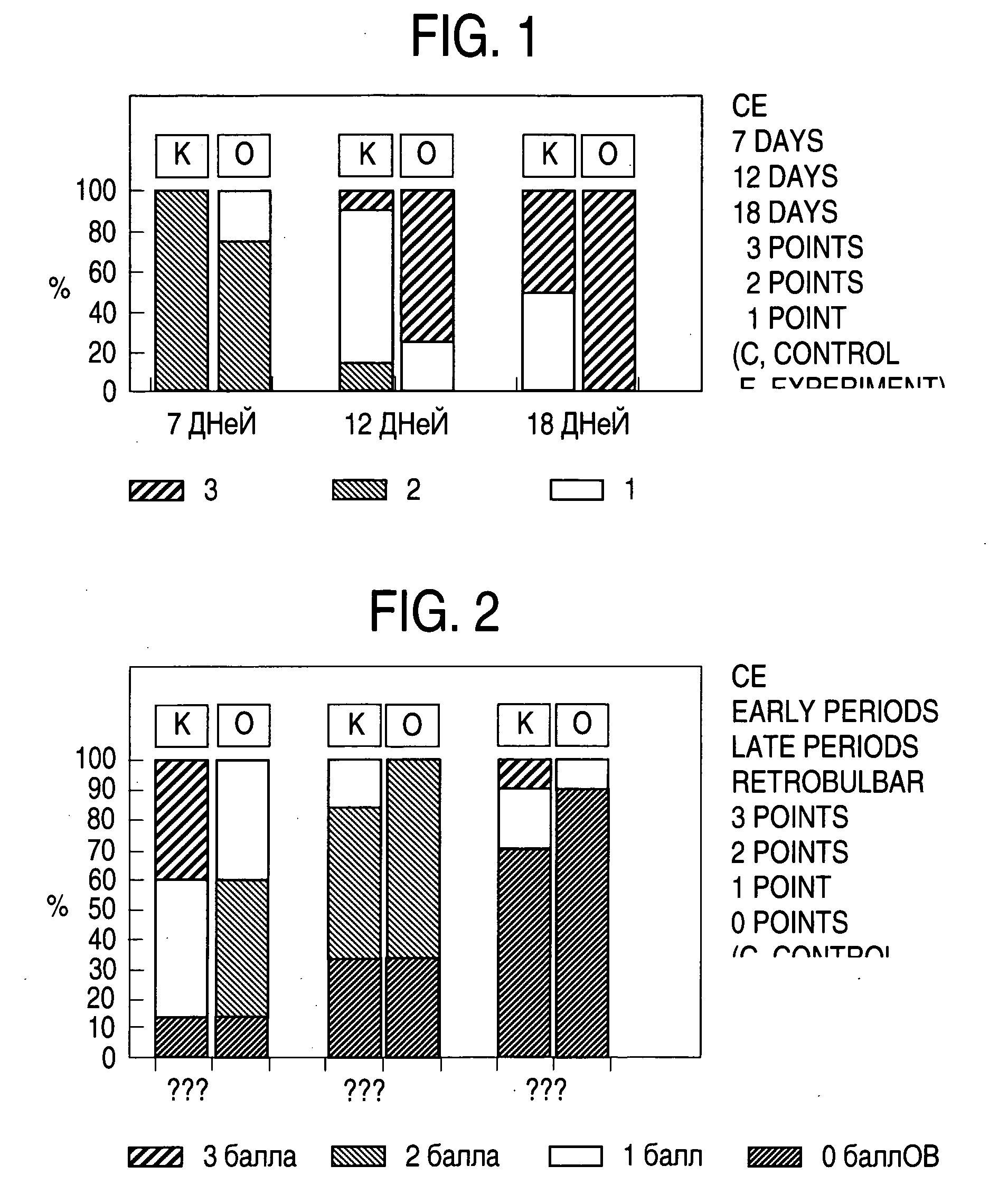
[0156]Chinchilla rabbits (body mass 2.5 to 3.0 kg, n=15) were used throughout the study. Two types of laser damage were induced (see the Table 5)

TABLE 5Experimental groupGroup 1Group 2Extent of laserSmall laser burns (noCoagulation of blood vesselsdamagedeliberate attempts(produced large areas ofwere made to targetischemic damage)blood vessels)Laser power300 milliWatt600-800 milliWattFocused beam200 micron300 micronspot sizeLaser pulse100 millisecond100 milliseconddurationNumber of burn300 spots per eye100 spots per eyespotsSpots locationLower quadrants of theRetinal blood vesselsfunduswere coagulated with the laseralong their entire length. Zoneof coagulation was inside acircle centered around opticdisk. Circle radius was halfthe equatorial radius of theeye.Number of5 rabbits10 rabbitsanimalsInjection sitesupra-choroid spacesupra-choroid ...

example 3

The Effect of Human Mesenchymal Cell Transplantation in the Treatment of Retina Laser Damage

Materials and Methods

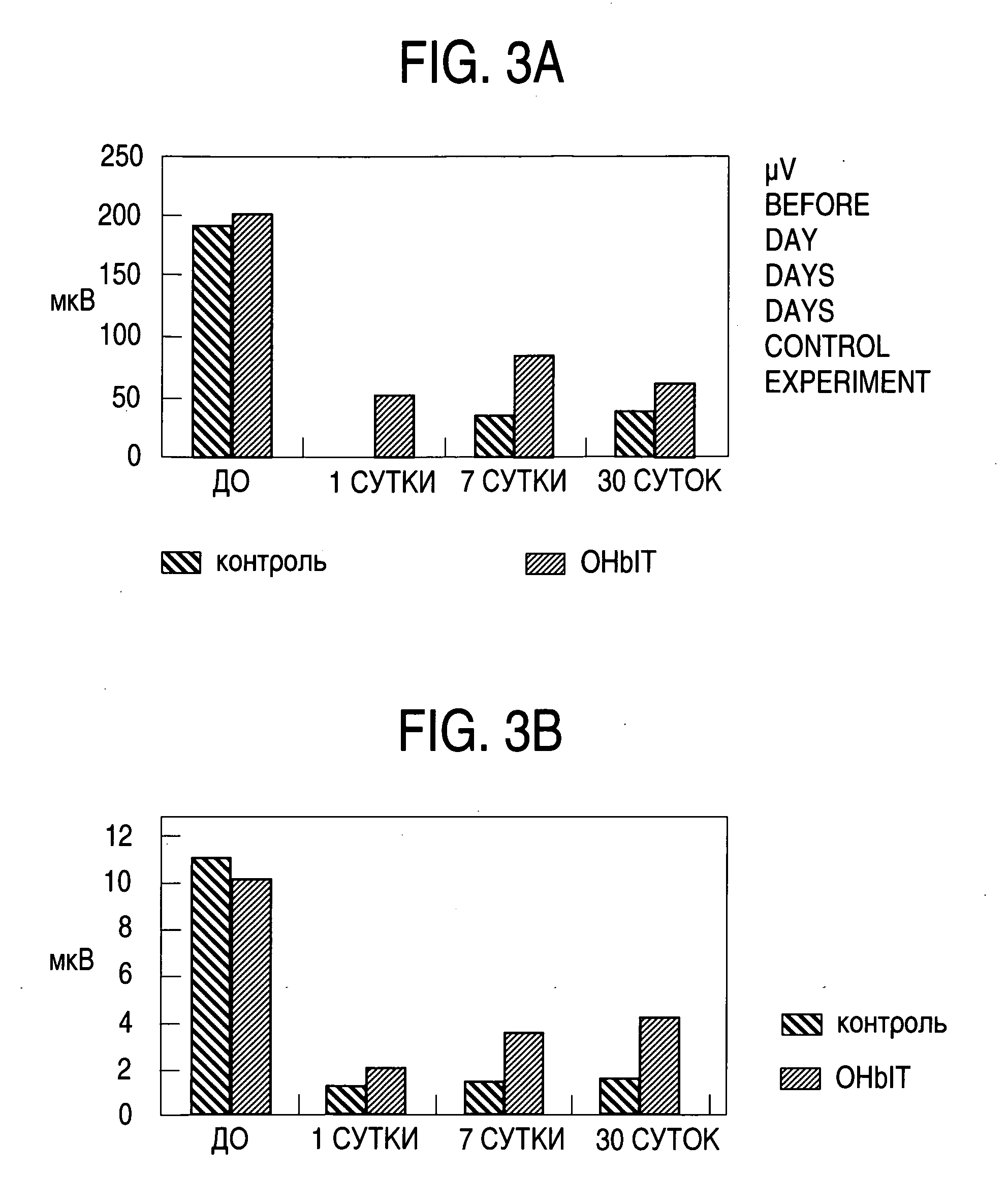
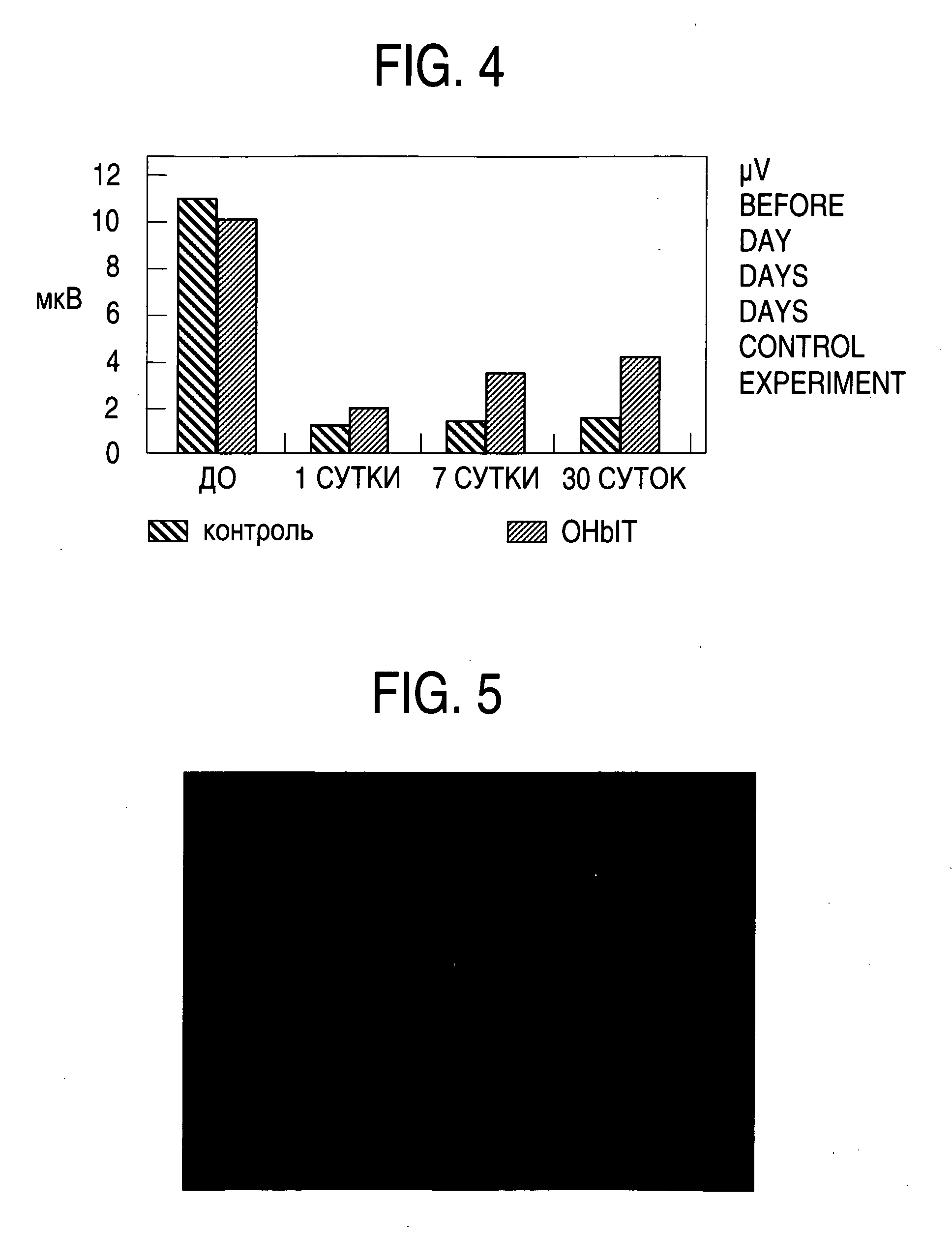
[0183]Chinchilla rabbits (body mass 2.5 to 3.0 kg, n=37) were used throughout the study. Laser-induced retinal damage was used as a model to simulate exudative macular degeneration in humans. Laser photocoagulation (200 μm spot size, 100 millisecond duration, 300 mW laser power ______ nm wavelength) was used to generate 300 laser spots in the lower quadrant(s) of each eye.

TABLE 11Animals were divided into 3 experimental groups:Exp.Exp.Exp.Group 1Group 2Group 3Number of human300,000 cells300,000 cells300,000 cellsmesenchymal cellsinjectedinjection siteinto theinto theinto thevitreous cavitysuprachoroidsuprachoroidspacespace (*)injection volume100 microL20 microL20 microLTime between human30 days30-40 days10-18 dayscell transplantationand animal euthanasiaMeasuring equipmentRetinoGrafRetinoGrafMjölner****ERG system Mjolner → http: / / www.globaleyeprogram.com / htm / mjolner.htm(*...


PUM


Abstract

A method for treating the side effects resulting from ophthalmic lasery surgery of the eye. The method administers stem cells as a means for treating the destruction of retinal and other tissues resulting from photocoagulation in the treatment of diabetic retinopathy and other eye disorders. Methods for using the invention with other ophthalmic laser surgical procedures, and other eye disorders, are also disclosed.

Description

PRIORITY[0001]This application claims priority from provisional application Ser. No. 61 / 038,711 filed May 30, 2008, the entire contents of which are incorporated herein by reference.FIELD OF THE INVENTION[0002]The invention relates to stem cell therapy. Specifically, the invention relates to the use of therapeutic stem cells in treating the damaging side effects of opthalmic laser surgery.BACKGROUND[0003]Photocoagulation, also called laser treatment, is a group of therapeutic procedures that combine the use of a laser, opthalmic devices and lenses to create tiny burns on the retina. Photocoagulation may assume different methodologies depending on the condition that is being treated.[0004]Focal macular laser therapy is one form of photocoagulation that is often used to treat disorders affecting the blood vessels of the macula, in particular diabetic retinopathy and retinal vein occlusion. The use of a laser in this application decreases the leakage from damaged blood vessels, helping...

Claims


Application Information

Patent Timeline
Patent Type & Authority Applications(United States)
IPC IPC(8): A61K35/14A61K35/12A61K35/28A61K35/30
CPCA61K35/30A61K35/28
Inventor TANKOVICH, NIKOLAI
Owner TANKOVICH NIKOLAI
Why Eureka
Try Eureka