Method and system for outlining a region in positron emission tomography studies

a positron emission tomography and region technology, applied in image enhancement, image analysis, instruments, etc., can solve the problems of individual images not optimal for analysis and visualization, pet images are typically characterized by a rather high level of noise, and the analysis of pet data is difficult to be independent, so as to achieve higher pixel intensity values and pixel intensity values

Inactive Publication Date: 2010-06-03
GE HEALTHCARE LTD
View PDF3 Cites 18 Cited by
  • Summary
  • Abstract
  • Description
  • Claims
  • Application Information

AI Technical Summary

Benefits of technology

[0024]According to an example embodiment of the present invention, a computer-readable medium may have stored thereon instructions adapted to be executed by a processor. The instructions, when executed, may cause the processor to perform a method for outlining a region of interest in a PET scan study. The method may provide for, during each of a plurality of scans of at least a part of a subject performed over a period of time that includes a plurality of sub-periods, generating corresponding data representing an image of the at least the part of the subject, where the image is defined by pixel intensities. Images represented by data generated during scans of different sub-periods may accentuate different features of the at least the part of the subject. The method may further provide for generating data representing an image having pixel intensities derived at least mostly from one or more images generated by the scanning of a single one of the sub-periods, determining which region of at least a portion of the image having the derived pixel intensities includes substantially higher pixel intensity values than other pixel intensity values of the at least a portion, and marking a peri

Problems solved by technology

Due to limitations in the amount of radioactivity administered to the subject, a usually short half-life of the radionuclide, and limited sensitivity of the recording system, PET images are typically characterized by a rather high level of noise.
This, together with a high level of non-specific binding to the target and sometimes small differences in target expression between healthy and pathological areas, are factors which make the analysis of PET data difficult independent of the utilized radionuclide or type of exper

Method used


Image

  • Method and system for outlining a region in positron emission tomography studies
  • Method and system for outlining a region in positron emission tomography studies
  • Method and system for outlining a region in positron emission tomography studies

Examples


Embodiment Construction

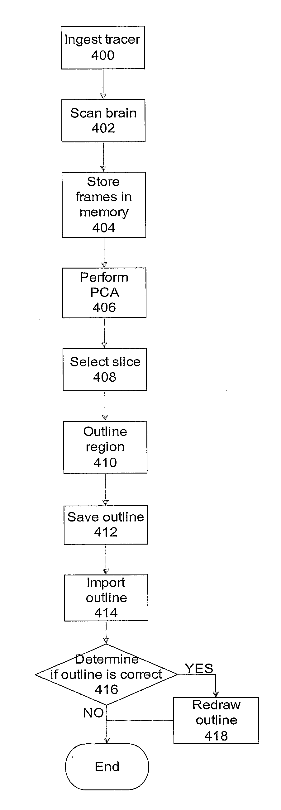
[0043]FIG. 3 is a block diagram that illustrates an example system according to an example embodiment of the present invention. A PET scanner 300 may be operated to generate multiple frames of a brain of a subject 301. Any suitably appropriate PET scanner may be used. For example, the scanner may be a Siemens ECAT HR+tomograph. The generated frames may be stored in a memory 310. The memory 310 may include any combination of conventional memory circuits, including electrical, magnetic, and / or optical systems. The memory 310 may include, for example, read only memory (ROM) 311, random access memory (RAM) 312, and / or bulk memory 313.

[0044]The memory 310 may have stored therein program instructions to be executed by a processor 305 for providing a modified PET image in which to outline a region of interest, e.g., a reference region, and / or for outlining the region of interest. The instructions may identify images in the memory 310 obtained from the scanner 300. For example, the image id...


PUM


Abstract

In a method and system for outlining a region of interest in a Positron Emission Tomography (PET) scan study, a processor may, based on application of Masked Volume Wise Principal Component Analysis (MVW-PCA) to a plurality of scan images, generate a PC2 image showing kinetic behavior of a particular part of a subject, in particular, the grey matter of the cerebellar cortex of the subject, and may outline, in the PC2 image, a region of the PC2 image having highest pixel intensity values of the PC2 image or of a portion thereof as a region of interest, and, in particular, as a reference region. The processor may generate a PC3 image showing kinetic behavior of a different part of the subject, in particular, blood vessels of the subject, import the outline into the PC3 image to determine the correctness of the outline, and modify the outline if it is incorrect.

Description

FIELD OF THE INVENTION[0001]The present invention relates to a method and system for outlining a region, in particular a reference region, in Positron Emission Tomography (PET) studies. The present invention further relates to a method and system for automatic outlining of the reference region.BACKGROUND INFORMATION[0002]PET is a conventionally known specialized imaging technique that uses tomography to computer-generate a three-dimensional image or map of a functional process in the body as a result of detecting photons when artificially introduced radionuclides (tracer) incorporated into biochemical substances decay and emit positrons. Analysis of the photons detected from the deterioration of these positrons is used to generate the tomographic images which may be quantified using a color scale to show the diffusion of the biochemical substances in the tissue indicating localization of metabolic and / or physiological processes. For example, radionuclides used in PET may be a short-...

Claims


Application Information

Patent Timeline
IPC IPC(8): G06K9/48G06T7/00
CPCG06T2207/10104G06T7/0083G06T7/12
Inventor RAZIFAR, PASHARINGHEIM, ANNALONGSTROM, BENGTENGLER, HENRYWALL, ANDERS
Owner GE HEALTHCARE LTD
Why Eureka
Try Eureka