Ultrasound imaging method and apparatus

Inactive Publication Date: 2010-06-24
GE MEDICAL SYST GLOBAL TECH CO LLC +1
View PDF21 Cites 29 Cited by
  • Summary
  • Abstract
  • Description
  • Claims
  • Application Information

AI Technical Summary

Benefits of technology

[0010]The embodiments described herein provide an ultrasound imaging method and apparatus, which

Problems solved by technology

However, there are also some drawbacks in ultrasound imaging, such as poor image contrast and repeatability.
The speckle noises may cover some useful information in the image and interfere the doctor's diagnosis to some extent.
Although spatial compounding reduces the noise speckles and optimizes the imaging quality to a certain degree, it still has some deficiencies.
So there is much complexity in implementation of this method.
The spatial compounding techniques in the prior art all combine successive image frames from different scan directions but with the same radiating/receiving frequency into one compounded image, and one common problem is that the selection of the steer angle of the ultrasonic beam is greatly limited by the transducer construction and emitting/receiving frequency, especially when happens to linear array probes with h

Method used


Image

  • Ultrasound imaging method and apparatus
  • Ultrasound imaging method and apparatus
  • Ultrasound imaging method and apparatus

Examples


Embodiment Construction

[0031]The embodiments of the present invention will be described in detail below with reference to the figures.

[0032]Linear scan is used as an example to illustrate the present invention in the following, but the present invention is not limited to linear scan, it can also be used in sector scan, etc.

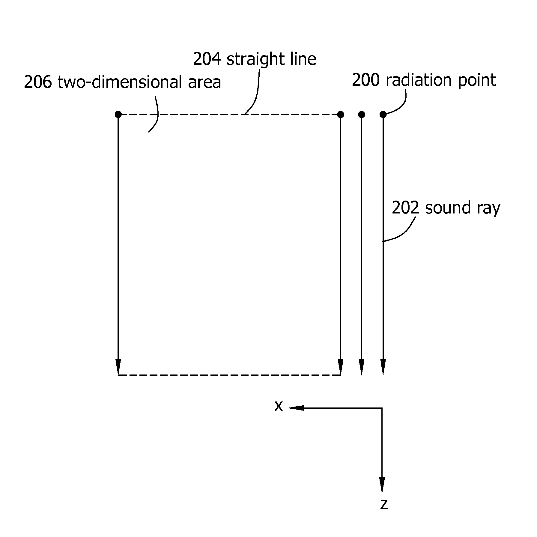
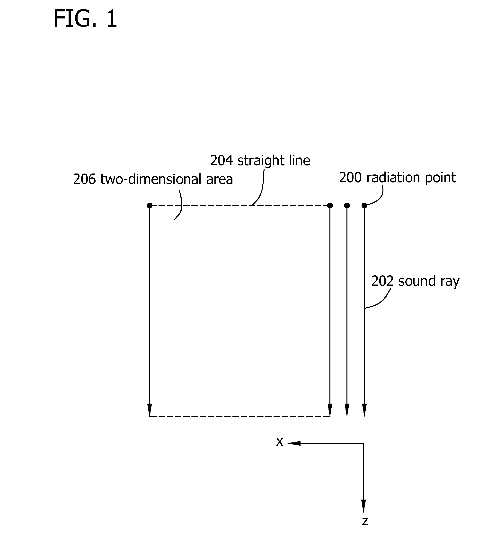
[0033]FIG. 1 illustrates the principle of scanning with a linear ultrasonic beam. As shown in FIG. 1, the scan is performed by a parallel excursion of a sound ray 202 along a straight line 204, said sound ray 202 is transmitted outside from a radiation point 200 in z direction, so that it sweeps a two-dimensional rectangular area 206 in x direction, thereby realizing linear scan. Wherein, the sound ray 202 corresponds to the central axis of the ultrasonic beam. By way of a parallel excursion of the aperture of the ultrasonic beam, scanning with the sound ray 202 in the scan direction is realized. By way of continuously changing the combination of a plurality of ultrasonic transducers wh...


PUM


Abstract

An ultrasound imaging method includes scanning an inspected object with a first cluster of ultrasonic beams having a first frequency and a first steer angle, to produce a first sub-frame which is used as a reference frame, and scanning the inspected object with a second cluster of ultrasonic beams having a second frequency different from the first frequency and a second steer angle different from the first steer angle, to produce a second sub-frame. The ultrasound imaging method also includes compounding the second sub-frame and the first sub-frame to form a compounded image, and displaying the compounded image.

Description

CROSS REFERENCE TO RELATED APPLICATIONS[0001]This application claims the benefit of Chinese Patent Application No. 200810184425.7 filed Dec. 19, 2008, which is hereby incorporated by reference in its entirety.BACKGROUND OF THE INVENTION[0002]The embodiments described herein relate to ultrasound imaging method and apparatus, in particular to ultrasound imaging method and apparatus using a technique for spatial and frequency compounding to obtain compounded ultrasound images.[0003]The diasonograph is a kind of ultrasound imaging apparatus, which images the inner structure of a human body by using ultrasonic waves (hereinafter referred to as “ultrasound”) as information carriers, and the image information thereof corresponds to the real structure of the human body in respect of the spatial and time distribution. Medical ultrasound imaging establishes images by the echo (the reflected wave or backscattered wave of the ultrasound by the human tissues) of different sound strengths produce...

Claims


Application Information

Patent Timeline
IPC IPC(8): A61B8/14
CPCA61B8/4209A61B8/0841A61B8/4281
Inventor HALMANN, MENACHEMFAN, HUACHEN, HUIREN
Owner GE MEDICAL SYST GLOBAL TECH CO LLC
Why Eureka
Try Eureka