

Method for inducing autophagy
- Summary
- Abstract
- Description
- Claims
- Application Information
AI Technical Summary
Benefits of technology
Problems solved by technology
Method used
Image

Examples
example 1
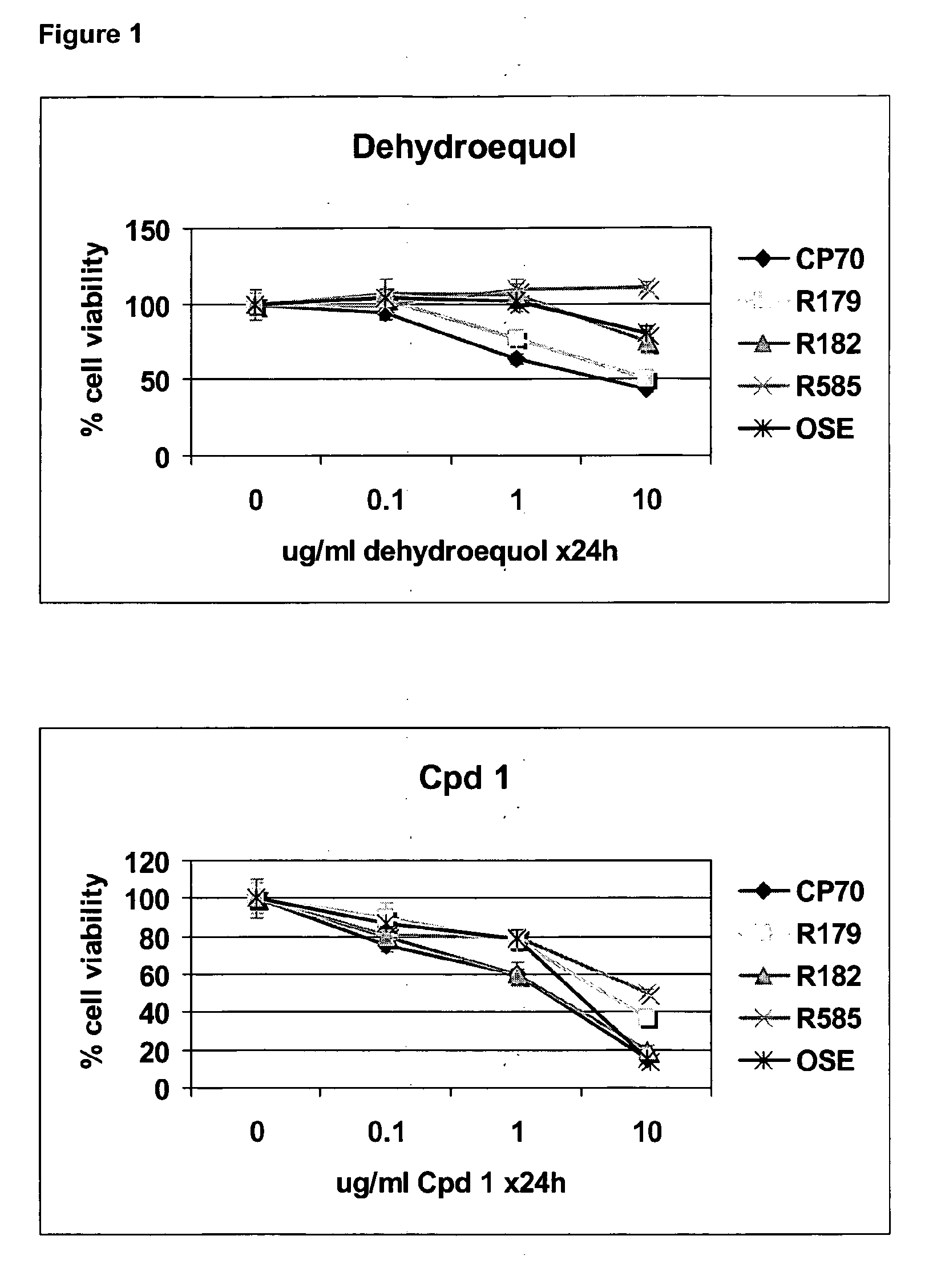
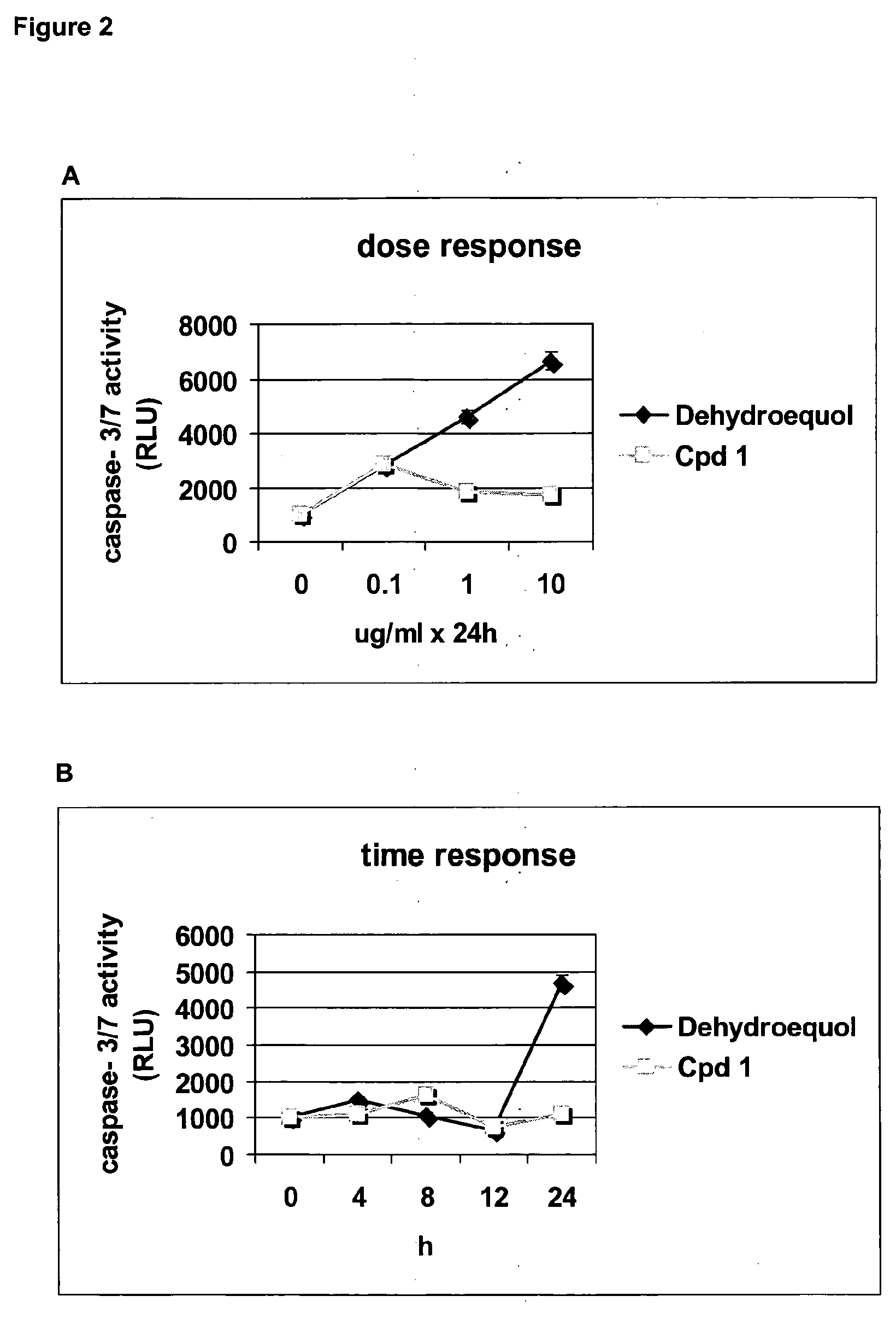
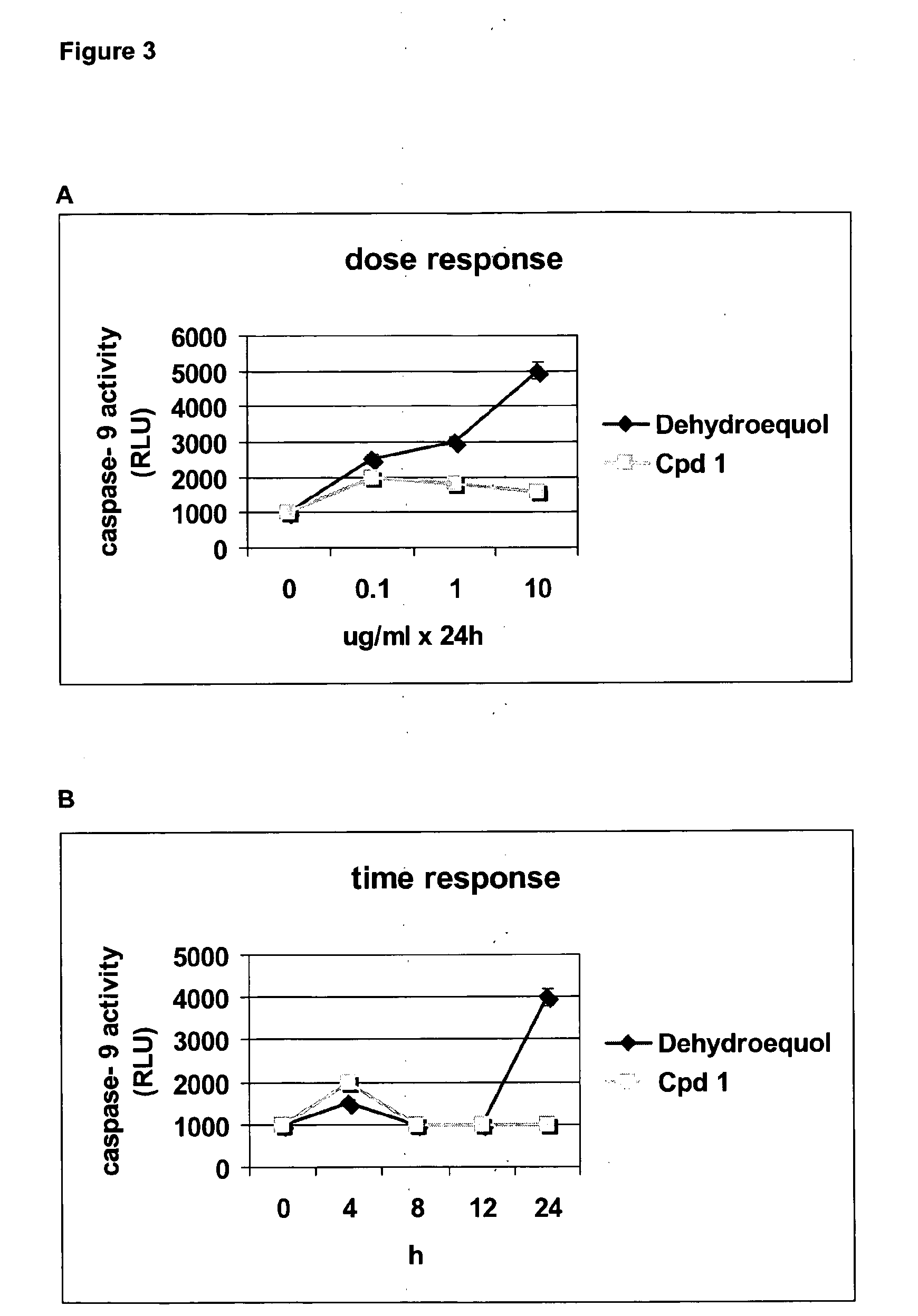
Cpd 1 Induces Cell Death Via a Caspase Independent Pathway
[0144]Human EOC cell lines A2780, CP70, and OSE were propagated in RPMI plus 10% fetal bovine serum (FBS) (Gemini Bio-Products, Woodland, Calif.) at 37° C. in a 5% CO2 atmosphere. Primary EOC cells (R179, R182, R585) were isolated from malignant ovarian ascites and cultured as previously described (Kamsteeg et al., 2003). Dehydroequol and Compound 1 (Cpd 1) were obtained from Novogen (Australia). All other reagents were purchased from Sigma Chemical (St. Louis, Mo.). The pan caspase inhibitor Z-VAD-FMK was obtained from R&D Systems (Minneapolis, Minn.).
[0145]Cell viability was evaluated using the CellTiter 96 Aqueous One Solution Cell Proliferation Assay (Promega, Madison, Wis.) according to the manufacturer's instructions. The values from the treated cells were compared with the values generated from the untreated cells and reported as percent viability. Each experiment was performed in triplicate.
[0146]For caspase activity ...
example 2
Vacuole Formation and DNA Fragmentation in the Presence of Cpd 1
[0149]R182 cells treated with Cpd 1 were observed microscopically to determine if morphological and structural alterations to cellular integrity took place. Cells treated with Cpd 1 were hyper-vacuolated (see FIG. 6) and the plasma membrane appeared to bleb or form folds. Phase-contrast images of Cpd 1-treated R182 cells treated with vehicle showed no evidence of vacuole formation, whereas in the presence of 5 μg / ml Cpd 1 over 4 hr and 8 hr vacuole formation is clearly evident (FIG. 6). These morphological changes observed in Cpd 1 treated cells are considered the hall mark of autophagy with the increased vacuoles formation thought to result in autophagosomes which eventually fuse with lysosomes to form autolysosomes. These structures contain catabolic hydrolases which are released into the cytoplasm when the lysosomal membrane becomes compromised.
[0150]The inventors then determined whether treatment with Cpd 1 induced ...
example 3
Protein Expression and Localisation Following Treatment with Cpd 1
[0151]After treatment with Cpd 1, protein was extracted from cells and measured as previously described (Kamsteeg et al., 2003). For separation of the cytoplasmic and mitochondrial fractions, cell pellets were processed using the ApoAlert Cell Fractionation kit (Promega) according to the manufacturer's instructions. 20 μg protein was denatured in sample buffer (2.5% sodium dodecyl sulfate [SDS], 10% glycerol, 5% β-mercapto-ethanol, 0.15 M Tris, pH 6.8, and 0.01% bromophenol blue) and subjected to 12% SDS polyacrylamide gel electrophoresis (SDS-PAGE) as previously described (Kamsteeg et al., 2003). The following antibodies and concentrations were used: mouse anti-Bax (BD Biosciences, 1:500), rabbit anti-actin (Sigma, 1:10,000), rabbit anti-cytochrome c (BD Biosciences, 1:1,000), mouse anti-Cox-4 (BD Biosciences, 1:500), rabbit anti-beclin 1 (Santa Cruz Biotechnology, 1:600), mouse anti LC3 (Nanotools, 1:500). These dil...
PUM
| Property | Measurement | Unit |
|---|---|---|
| Cell death | aaaaa | aaaaa |
| Stability | aaaaa | aaaaa |
Abstract
Description
Claims
Application Information