
Method for percutaneous lateral access to the left ventricle for treatment of mitral insufficiency by papillary muscle alignment

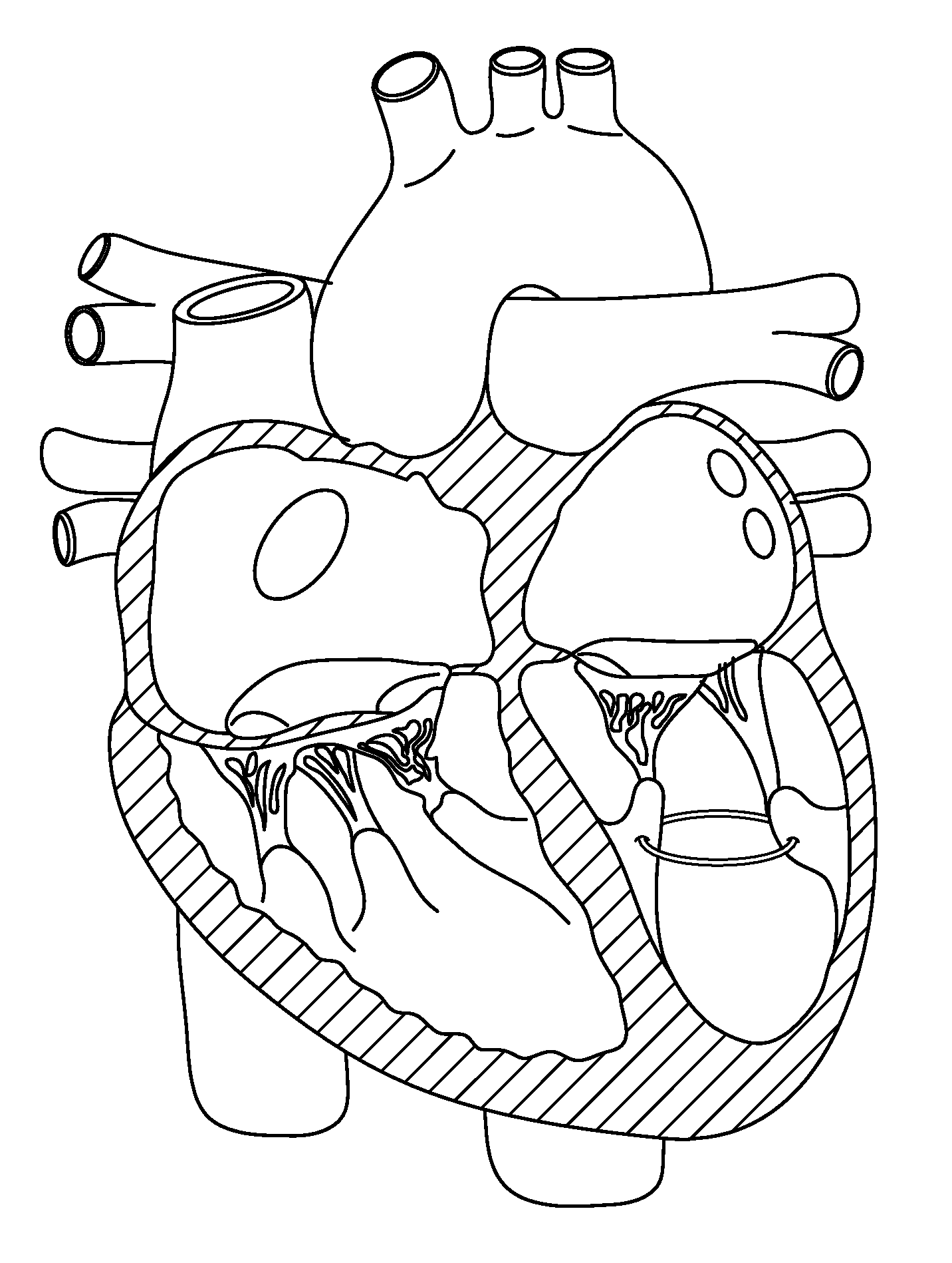
a technology of papillary muscle and lateral access, which is applied in the field of therapeutic changing the geometry of the left ventricle of the human heart, can solve the problems of ventricular tissue damage, dilated left ventricle, and insufficient heart pumping or other heart function abnormalities, so as to improve cardiac function and reduce the volume of the left ventricle

Inactive Publication Date: 2010-08-19
TENDYNE MEDICAL
View PDF99 Cites 143 Cited by
  • Summary
  • Abstract
  • Description
  • Claims
  • Application Information

AI Technical Summary

Benefits of technology

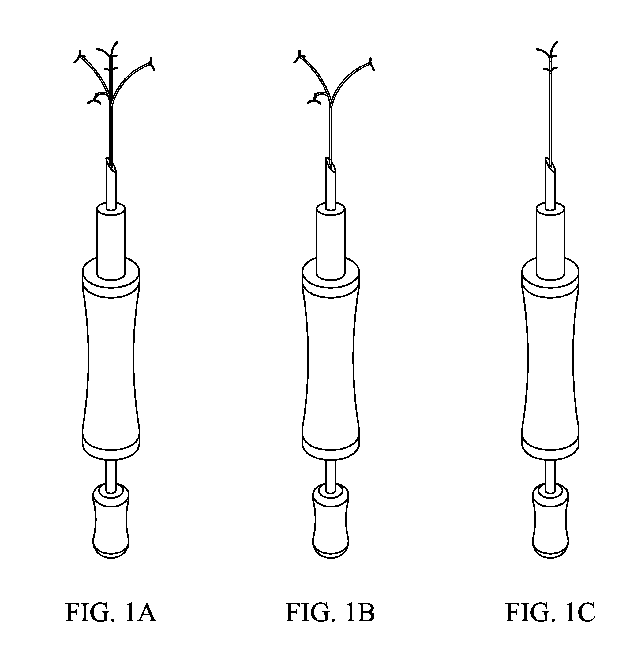
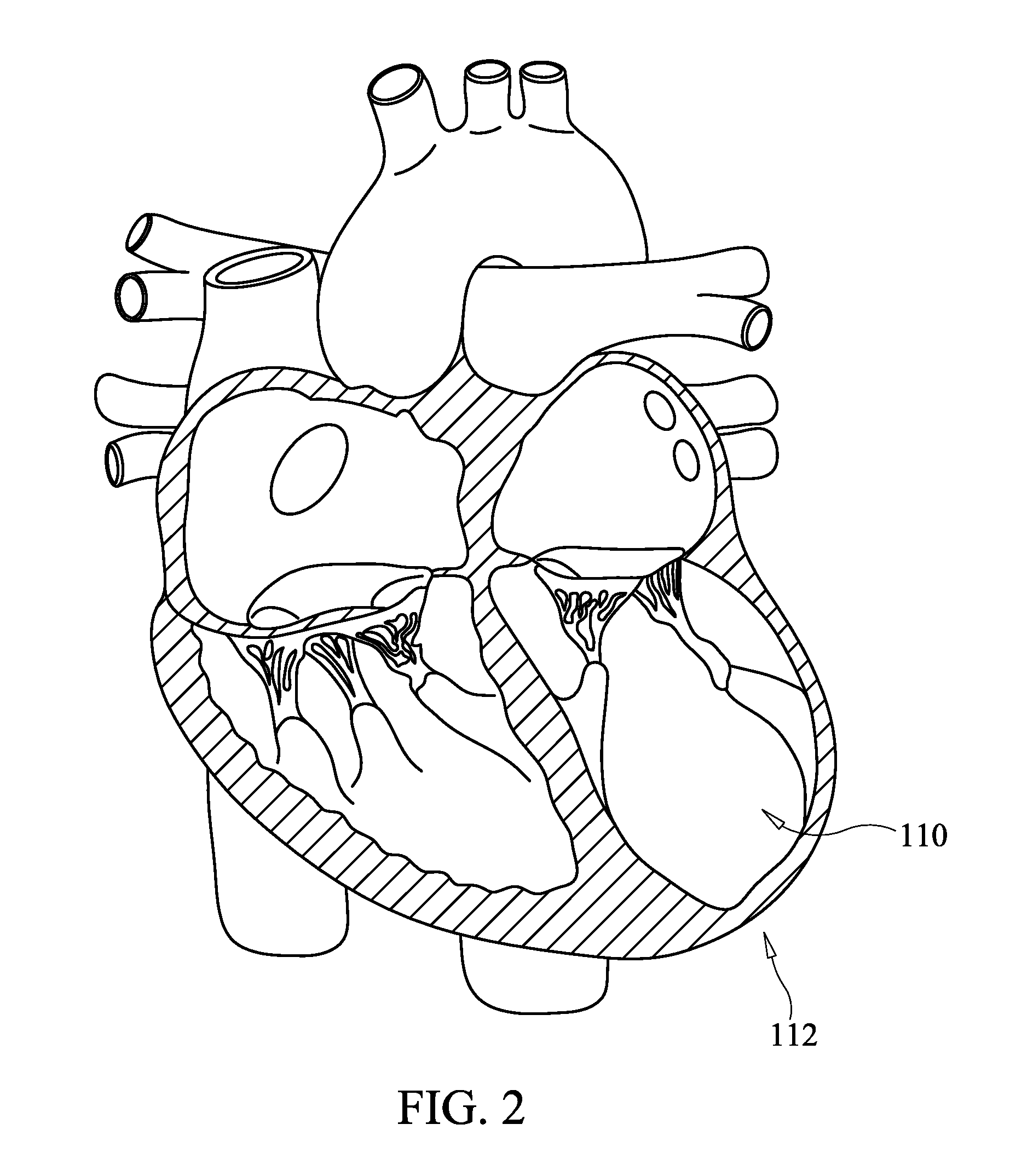
[0018]Accordingly, in a preferred embodiment of the invention, there is provided a method for improving cardiac function, comprising the steps of: inserting by percutaneous approach a tether installation device into a patient; and inserting said tether installation device through the lateral wall of the left ventricle of the patient's heart; attaching at least one tethered papillary muscle anchor from within said tether installation device to a papillary muscle within said left ventricle; withdrawing said tether installation device from said left ventricle such that the tether of the tethered papillary muscle anchor tranverses the left ventricular wall and extends from the inside to the outside of the left ventricle; attaching a pledget to the tether portion outside the left ventricle to form a wall anchor; wherein said papillary anchor and said wall anchor are joined by the tether to change the geometry and reduce the volume of the left ventricle.
[0019]In another preferred embodiment of the invention, there is provided a method for reducing ventricular volume, comprising the steps of : inserting by percutaneous approach a tether installation device into a patient; and inserting said tether installation device through the lateral wall of the left ventricle of the patient's heart and into the left ventricle of the patient's heart; and attaching a first tethered papillary muscle anchor from within said tether installation device to a papillary muscle within said left ventricle; attaching a second tethered papillary muscle anchor from within said tether installation device to a second papillary muscle of the left ventricle of the patient's heart; withdrawing said tether installation device from said left ventricle such that the tethers of the tethered papillary muscle anchors tranverse the left ventricular wall and extend from the inside to the outside of the left ventricle; attaching one or more pledgets to the tether portions outside the left ventricle to form one or more wall anchors; wherein said papillary anchors and said wall anchor are joined by the tethers to change the geometry and reduce the volume of the left ventricle.
[0031]In another preferred embodiment, the needle is a non-coring needle to reduce defect to the tissue(s). Preferably, the needle used is a small gauge needle used as a guide to reduce the defect made to the tissue, and the opening is then temporarily dilated using the larger bore instrument to house the catheter.
[0033]In another preferred embodiment of the invention, there is provided a medical device for improving cardiac function or reducing ventricular volume, comprising: a canula having a tethering device disposed therein; said canula having a trocar or needle for percutaneously accessing the chest cavity by intercostal or subxyphoid introduction, said trocar or needle capable of piercing the lateral wall of the left ventricle of the patient's heart and a leakage control hemostasis valve / sleeve; said tethering device comprising at least one first papillary muscle anchor for attaching to a first papillary muscle within said left ventricle and at least one second papillary muscle anchor for attaching to the second papillary muscle of the left ventricle of the patient's heart; said tethering device further comprising a tether member for joining said first papillary muscle anchor to said second papillary muscle anchor so as to reduce the left ventricular volume of the patient.

Problems solved by technology

According to the Center for Disease Control, heart disease is the leading cause of death in the United States and is a major cause of disability.
It often results in inadequate heart pumping or other heart function abnormalities.
However, a dilated left ventricle is generally due to the effects of a myocardial infarction.
This downstream ventricular tissue is damaged, but since the volume of blood that fills the ventricle does not change, the damaged organ has to work harder to eject the blood.
This increased load causes an increase in the radius of the ventricle and the thickness of the ventricular wall changes.
Overall, the dilated left ventricle cannot produce a strong enough pulse to maintain health and efficient circulatory return.
The disadvantage to the Dor procedure is that it places synthetic tissue inside the LV cavity and it is usually done as part of a coronary artery bypass graft (open heart) surgery.
However, cardiac catheterization poses the risk of blood clots that can trigger strokes, damage to blood vessels, and damage to the heart or pericardium.

Method used


Image

  • Method for percutaneous lateral access to the left ventricle for treatment of mitral insufficiency by papillary muscle alignment
  • Method for percutaneous lateral access to the left ventricle for treatment of mitral insufficiency by papillary muscle alignment
  • Method for percutaneous lateral access to the left ventricle for treatment of mitral insufficiency by papillary muscle alignment

Examples


Embodiment Construction

Definitions

[0052]The following definitions are provided as an aid to understanding the detailed description of the present invention.

[0053]“Anchors” for the purposes of this application, is defined to mean any fastener. Thus, anchors may comprise C-shaped or semicircular hooks, curved hooks of other shapes, straight hooks, barbed hooks, clips of any kind, T-tags, or any other suitable fastener(s). In one embodiment, anchors may comprise two tips that curve in opposite directions upon deployment, forming two intersecting semi-circles, circles, ovals, helices or the like. In some embodiments, anchors are self-deforming. By “self-deforming” it is meant that anchors change from a first undeployed shape to a second deployed shape upon release of anchors from restraint in housing. Such self-deforming anchors may change shape as they are released from housing and enter papillary or myocardial tissue, to secure themselves to the tissue. Thus, a crimping device or other similar mechanism is ...


PUM


Abstract

This invention relates to devices and methods for the therapeutic changing of the geometry of the left ventricle of the human heart. Specifically, the invention relates to the left-ventricular lateral wall introduction of an anchoring device to align the papillary muscles.

Description

CROSS REFERENCE TO RELATED APPLICATIONS[0001]This application is a continuation-in-part and claims priority under 35 USC 120 to U.S. Ser. No. 12 / 691,591, filed 21 Jan. 2010, entitled Apical Papillary Muscle Attachment for Left Ventricular Reduction, the contents of which are incorporated by reference herein in their entirety, which claims priority benefit under 35 USC 119(e) to U.S. 61 / 146,144, filed 21 Jan. 2009.STATEMENT REGARDING FEDERALLY SPONSORED RESEARCH OR DEVELOPMENT[0002]No federal government funds were used in researching or developing this invention.NAMES OF PARTIES TO A JOINT RESEARCH AGREEMENT[0003]n / aREFERENCE TO A SEQUENCE LISTING[0004]n / a.BACKGROUND[0005]1. Field of the Invention[0006]This invention relates to devices and methods for the therapeutic changing of the geometry of the left ventricle of the human heart. Specifically, the invention relates to the percutaneous lateral introduction of an anchoring device to align the papillary muscles.[0007]2. Background of...

Claims


Application Information

Patent Timeline
IPC IPC(8): A61F2/00
CPCA61B17/00234A61B17/0401A61F2/2487A61B2017/048A61F2/2457A61B17/0487
Inventor SCHANKERELI, KEMAL
Owner TENDYNE MEDICAL
Learn More