
Blood examination apparatus

a blood examination and apparatus technology, applied in the field of blood examination apparatus, can solve the problems of difficult to find cancer cells in a large amount of blood, easy to block the physical mesh, and inability to cytometry, etc., and achieve the effect of high speed

Active Publication Date: 2010-09-02
HAMAMATSU PHOTONICS KK
View PDF15 Cites 2 Cited by
  • Summary
  • Abstract
  • Description
  • Claims
  • Application Information

AI Technical Summary

Benefits of technology

[0030]According to the present invention, identification and position identification of cancer cells in blood can be performed at a high speed.

Problems solved by technology

However, this physical mesh is easily clogged.
In actuality, it has been attempted to detect cancer cells in blood by using flow cytometry (refer to Non-Patent Documents 7 to 9), however, the flow cytometry is not an apparatus intended to examine as much blood as 5000 mL.
Therefore, it is not realistic to find cancer cells in a large amount of blood by analyzing images taken by a CCD camera or a CMOS camera.
However, this technique also requires analysis of the images taken by the CCD camera, and therefore, it is not realistic to find cancer cells in a large amount of blood.

Method used


Image

  • Blood examination apparatus
  • Blood examination apparatus
  • Blood examination apparatus

Examples


Embodiment Construction

[0039]Hereinafter, best modes for carrying out the present invention will be described in detail with reference to the accompanying drawings. In the description of the drawings, the same components are designated by the same reference numerals and letters, and overlapping description will be omitted.

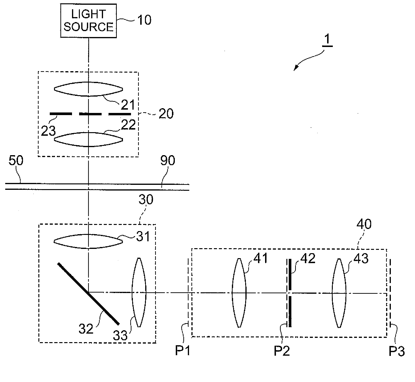
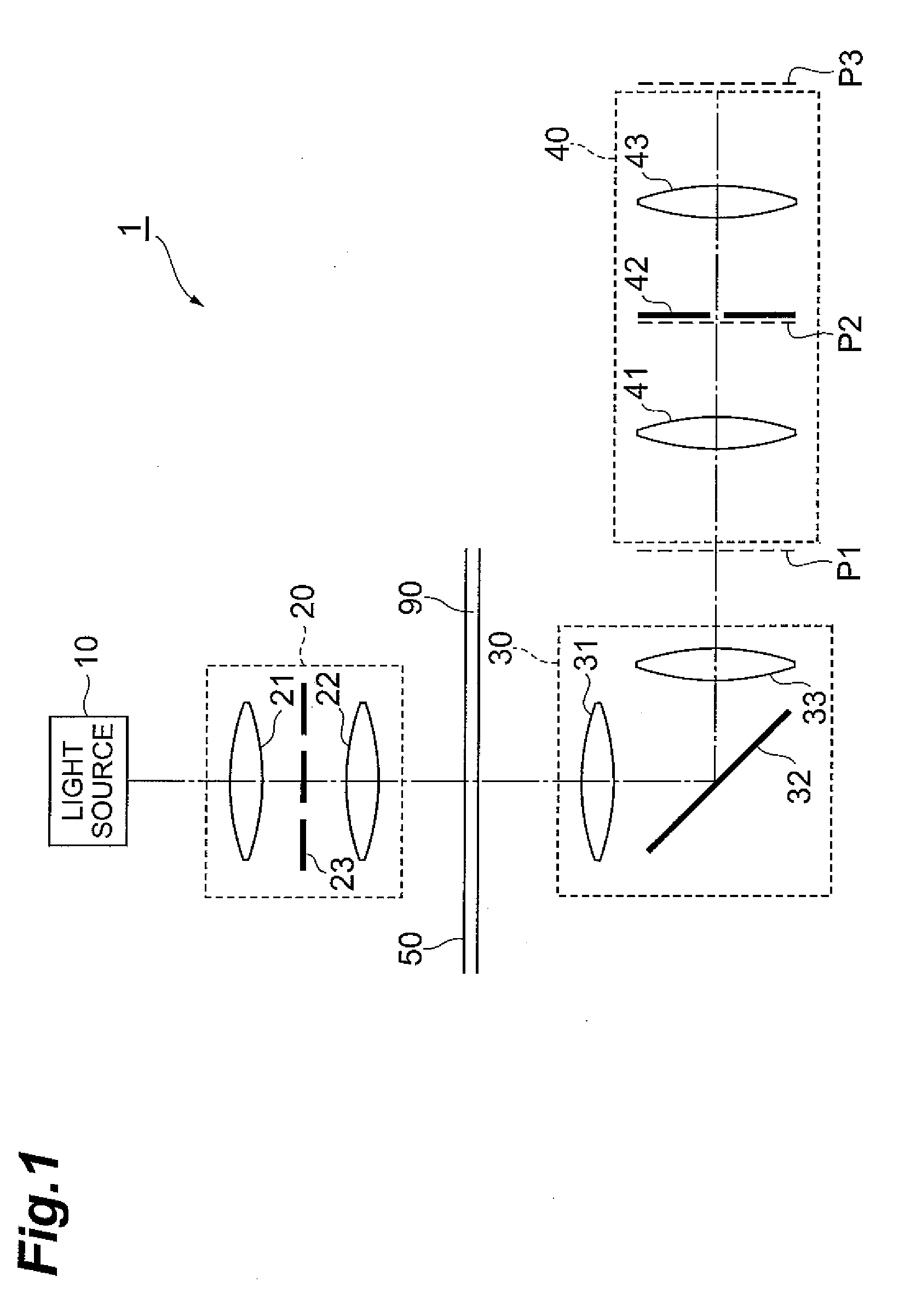
[0040]FIG. 1 is a configuration view of a blood examination apparatus 1 according to the present embodiment. The blood examination apparatus 1 shown in this figure includes a light source 10, an irradiation optical system 20, an imaging optical system 30, a detection optical system 40, and a flow cell 50. Among these, the light source 10, the irradiation optical system 20, and the imaging optical system 30 have the same configurations as those of a phase-contrast microscope. The detection optical system 40 includes a first Fourier transformation optical system 41, a spatial light filter 42, and a second Fourier transformation optical system 43. The flow cell 50 may be a blood vessel or a...


PUM

PropertyMeasurementUnit
sizeaaaaaaaaaa
sizeaaaaaaaaaa
sizeaaaaaaaaaa

Abstract

This blood examination apparatus examines cancer cells mixed in an examination object which is flowing blood, and includes: a flow cell through which the examination object is made to flow; an imaging optical system which light output from the examination object in an examination region in the flow cell enters, the imaging optical system forming an image of the light on a first image plane; a first Fourier transformation optical system which optically two-dimensionally Fourier-transforms the image formed on the first image plane by the imaging optical system to form the Fourier-transformed image on a second image plane; a spatial light filter which selectively allows a portion in a certain range around an optical axis of the first Fourier transformation optical system of the image formed on the second image plane by the first Fourier transformation optical system to pass through; and a second Fourier transformation optical system which optically two-dimensionally Fourier-transforms the portion which has passed through the spatial light filter of the image formed on the second image plane by the first Fourier transformation optical system to form the Fourier-transformed image on a third image plane.

Description

TECHNICAL FIELD[0001]The present invention relates to an apparatus for examining blood without staining.BACKGROUND ART[0002]In recent years, in the medical and biotechnology fields, circulating tumor cells which are cancer cells invading blood from cancer tissue have attracted attention (refer to Non-Patent Document 1). That is, as one of the cancer metastasis mechanisms, there is hematogenous dissemination in which cancer cells developed in a certain organ are transported by blood and metastasize to other organs. It has been expected that metastasis of cancer can be suppressed by removing cancer cells in blood. It is hoped that if living cancer cells can be captured from blood, this will contribute to the development of medicines as antibodies, and the properties and mechanisms of metastasis of cancer will be unraveled.[0003]Blood cells in blood are briefly classified into red blood cells and white blood cells. Approximately 5×10⁹ red blood cells are contained in 1 mL of whole bloo...

Claims


Application Information

Patent Timeline
Patent Type & Authority Applications(United States)
IPC IPC(8): G01N33/48G06K9/00
CPCG01N15/1434G01N15/147G06T2207/30024G06T2207/20056G06T7/0012G01N2015/145G01N33/4915G01N2015/1493G02B21/14G01N2015/1006G01N15/1459G01N15/1433G01N15/149
Inventor IWAI, HIDENAOYAMAUCHI, TOYOHIKO
Owner HAMAMATSU PHOTONICS KK
Learn More