
Glaucoma shunts with flow management and improved surgical performance

a glaucoma and flow management technology, applied in the field of eye surgery, can solve the problems of affecting the vision of the afflicted eye, excessive buildup of aqueous fluid in the eye, and prohibitively expensive pharmacological treatment of the large majority of glaucoma patients, so as to improve the glaucoma shunt, reduce post-op complications, and effectively regulate the intraocular pressur

Active Publication Date: 2010-09-30
JOHNSON & JOHNSON SURGICAL VISION INC
View PDF68 Cites 127 Cited by
  • Summary
  • Abstract
  • Description
  • Claims
  • Application Information

AI Technical Summary

Benefits of technology

[0015]The present application provides various solutions for improving glaucoma shunts and reducing post-op complications. In a preferred embodiment, a glaucoma shunt having a pliable plate attached to the sclera and a drainage tube that extends into the inner eye causes a bleb to form around the plate which effectively regulates intraocular pressure (IOP). The pliable plate has a reduced size and may be microtextured in particular areas to control cell adhesion thereto. A flow restrictor within the drainage tube helps prevent excessive pressure buildup or dropoff immediately post-op, and an anti-microbial coating reduces the chance of infection.

Problems solved by technology

This increase in pressure is most commonly caused by stenosis or blockage of the aqueous outflow channel, resulting in excessive buildup of aqueous fluid within the eye.
This increase in IOP produces gradual and permanent loss of vision in the afflicted eye.
Pharmacological treatment is prohibitively expensive to a large majority of glaucoma patients.
In addition, many people afflicted with the disease live in remote or undeveloped remote areas where the drugs are not readily accessible.
The drugs used in the treatment, in particular steroids, often have undesirable side effects and many of the long-term effects resulting from prolonged use are not yet known.
Additionally, patient compliance is an issue and, for example, patients often forget their medication.
While often successful, these surgical techniques possess inherent risks associated with invasive surgery on an already afflicted or compromised eye.
Furthermore, the tissue of the eye can scar over this small area and the eye reverts to the pre-operative condition, thereby necessitating the need for further treatment.
Many problems occurred with the early Molteno device.
Further, this early device often did not reduce the IOP enough to treat the glaucoma without the use of additional medications.
The long tube Molteno implant is disadvantageous as the plates are formed of a rigid plastic which makes insertion beneath the eye tissue difficult and time-consuming.
An open drainage tube may release too much of the fluid too fast, which is detrimental to the eye.
However, such valved devices sometimes clog later, requiring another surgery.
This method works well, but the timing of suture dissolution is necessarily inexact, and a second procedure undesirable.
Though a bleb is thought to have a beneficial function in regulating aqueous humor diffusion, too large of a bleb may cause the patient some pain or may be aesthetically unacceptable.
This complication may occur in any intraocular surgery, with possible loss of vision and the eye itself.
The risk of infection is more pronounced early in a glaucoma shunt procedure when a passage to the interior of the eye is created and fluid flows therethrough.

Method used


Image

  • Glaucoma shunts with flow management and improved surgical performance
  • Glaucoma shunts with flow management and improved surgical performance
  • Glaucoma shunts with flow management and improved surgical performance

Examples


Embodiment Construction

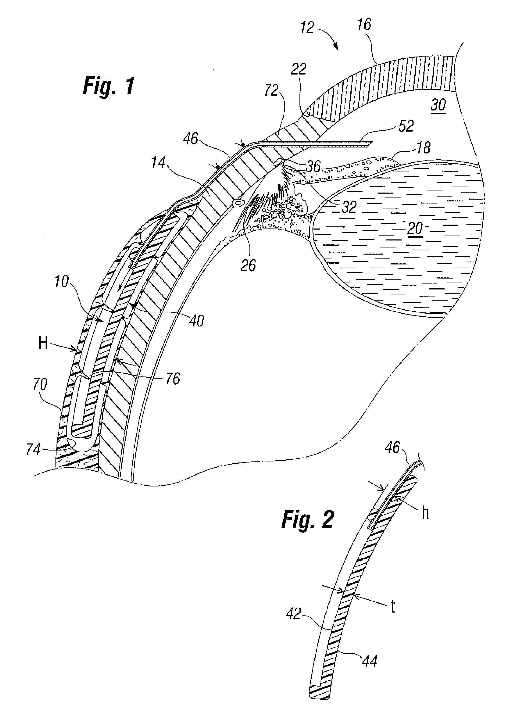
[0054]FIG. 1 illustrates a glaucoma shunt 10 constructed in accordance with the present application positioned within the tissue of an eye 12 (looking upward relative to the page). The relevant structures of the eye 12 will be described briefly below to provide background for the anatomical terms incorporated herein, however, it should be realized that a number of anatomical details have been omitted for clarity of understanding. The tough outer membrane known as the sclera 14 covers all of the eye 12 except that portion covered by the cornea 16, the thin, transparent membrane which enables light to enter the pupil 18 defined by the iris aperture in front of the lens 20. The cornea 16 merges into the sclera 14 at a juncture referred to as the limbus 22. The ciliary body 26 begins near the limbus 22 and extends along the interior of the sclera 14.

[0055]It is well-known that aqueous humor is produced by the ciliary body 26 and reaches the anterior chamber 30 formed between the iris 18...


PUM


Abstract

A method of treating glaucoma in an eye by managing fluid flow past an implanted shunt having an elastomeric plate and a non-valved elastomeric drainage tube. The plate is positioned over a sclera of the eye with an outflow end of the elastomeric drainage tube open to an outer face of the plate. An inflow end of the drainage tube tunnels through the sclera to the anterior chamber of the eye. The plate may have regions of greater propensity for cell adhesion alternating with regions of lesser cell adhesion. For example, regions of texturing around the plate or drainage tube may be provided to control the size of a bleb that forms over the implant. The effective surface area of the plate may be balanced against a number of fenestrations. The drainage tube has a reduced profile and may be shaped with a non-circular external cross-section to reduce its height. A scleral groove may be used to further reduce the height of the drainage tube on the sclera. A flow restrictor for the early post operative period will immediately lower the intraocular pressure (IOP) and simultanously prevent hypotony.

Description

RELATED APPLICATIONS[0001]The present application claims priority under 35 U.S.C. §119(e) to Provisional Application No. 61 / 163,862, filed on Mar. 26, 2009.FIELD OF THE INVENTION[0002]The present invention relates to treatments for glaucoma in the eye and, more particularly, to glaucoma shunts and methods of use for draining fluid from the eye at a controlled rate.BACKGROUND OF THE INVENTION[0003]Intraocular pressure in the eye is maintained by the formation and drainage of aqueous humor, a clear, colorless fluid that fills the anterior and posterior chambers of the eye. Aqueous humor normally flows from the anterior chamber of the eye out through an aqueous outflow channel at a rate of 2 to 3 microliters per minute.[0004]Glaucoma is a progressive disease of the eye characterized by a gradual increase of intraocular pressure (IOP). This increase in pressure is most commonly caused by stenosis or blockage of the aqueous outflow channel, resulting in excessive buildup of aqueous fluid...

Claims


Application Information

Patent Timeline
IPC IPC(8): A61F9/00
CPCA61F9/00781
Inventor VAN DER MOOREN, MARRIE H.BOGAERT, THEOPHILUSJAIN, RAKHI
Owner JOHNSON & JOHNSON SURGICAL VISION INC
Learn More