While the treatment of silent seizures (with standard anti-epileptic drugs) is rather efficient, their detection remains the weakest point in the acute care of patients; they are impossible to detect upon standard clinical examination, and can only be revealed if the electrical activity of the brain is recorded (e.g., using an electroencephalographic (EEG) recorder).
EEG is however infrequently recorded in most neuro-
intensive care units because of the difficulties in using relatively bulky EEG systems in a crowded setting of an ICU, and in maintaining good electrical contact between the
EEG electrodes and the
scalp of the patient over longer periods of time.
The second approach is still largely limited to experimental settings, but with further improvements in computational technologies and
artificial intelligence it may find its way to clinicians.
Decreased CBF can cause a secondary,
ischemic brain injury, that is often more severe and extensive than the primary injury inflicted in an accident.
However,
standard methods for measuring CBF are technically complex, financially costly, and are applied in specialized institutions where the equipment and knowledge are available.
However, these methods are focused on only measuring the flow through the arterial portion of the cerebral vasculature.
TABLE 1Methods for measurement of CBFMethodsShortcommings for in-field useNitrous
oxide inhalationCumbersome, unsuitable for dynamicmethodchangesO215
positron emissionRadioactive, expensive, not portabletomography (PET)Single
photon emissionRadioactive, expensive, not portabletomography (SPECT)
Perfusion-weighted MRIExpensive, not portableXe-enhanced computerizedRadioactive, expensive, not portabletomography (CT)
Transcranial Doppler (TCD)Considerable expertise requiredIntracranial probes (basedInvasive, risk of infections, noton
laser flowmetry)
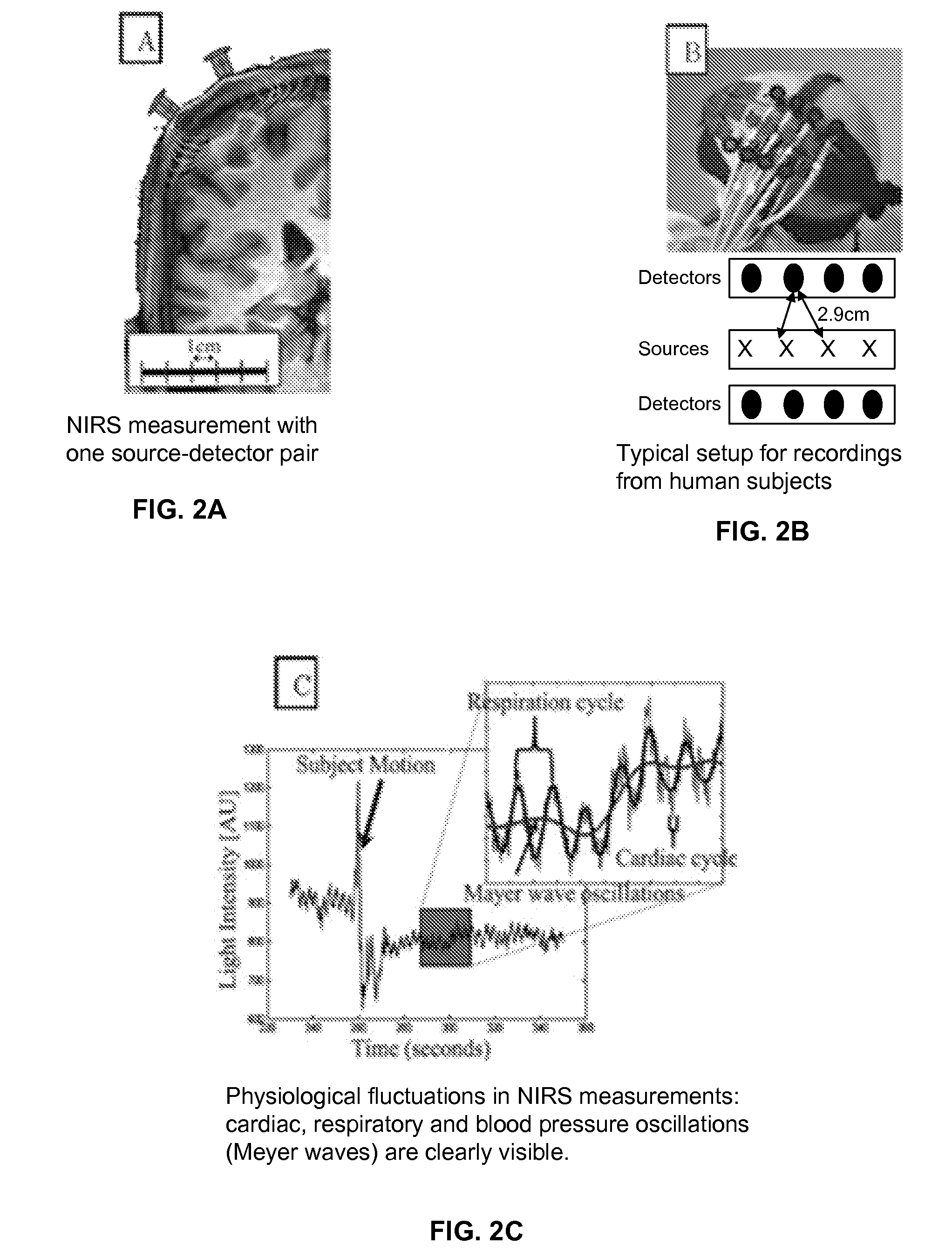
absolute measurementHowever, since the near-
infrared light penetrates only 5-10 mm into the head, NIRS can only monitor
blood flow in the
brain cortex but not in the deeper structures.
Other methods such as subarachnoid and epidural transducers or spinal tap have much lower accuracy.
However, the invasive methods for monitoring ICP share several common drawbacks: the transducers have to be calibrated before
insertion; their output drifts, requiring either a recalibration or replacement of the
catheter after 36-48 hours;
insertion of a
catheter carries a risk of brain or
spinal cord damage and infection; and the placement of a
catheter requires a highly trained individual, such as a neurosurgeon.
For these reasons, invasive ICP monitoring techniques cannot be used outside of the
hospital setting.
Table 2 illustrates that the non-invasive methods' common drawback is insufficient accuracy: the margins of error of ICP estimates are of the same
order of magnitude as the whole range of ICP that is clinically of interest (0-50 mmHg).

