Detection of uterine leiomyosarcoma using lmp2
a technology of uterine leiomyosarcoma and detection method, which is applied in the field of detection method of uterine leiomyosarcoma, can solve the problems of difficult differentiation of uterine leiomyoma from uterine leiomyosarcoma, and the differentiation is not always accura
- Summary
- Abstract
- Description
- Claims
- Application Information
AI Technical Summary
Benefits of technology
Problems solved by technology
Method used
Image

Examples
example 1
LMP2 Transcription and Expression in Human Uterine Leiomyosarcoma
[0149]In this example, the materials and methods described below were used.
Cell Strain and Medium
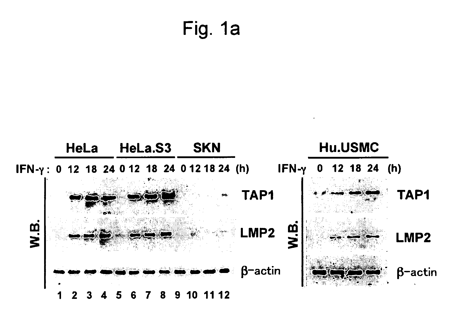
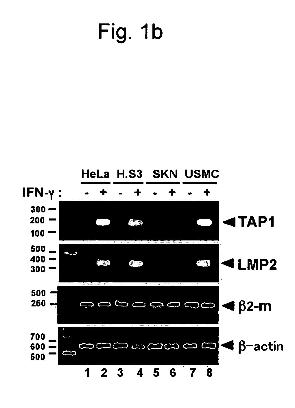
[0150]Human uterine leiomyosarcoma cell lines, i.e., SKN cells (RCB0513), were purchased from Cell Bank, RIKEN Bio Resource Center, and the cells were retained in F-12 Nutrient Mixture (Ham) medium (Invitrogen) supplemented with 0.6% L-glutamine (Invitrogen) and 15% fetal bovine serum (Sigma-Aldrich, Inc.). HeLa cells and HeLa.S3 cells were retained in Dulbecco's MEM supplemented with 0.6% L-glutamine and 10% fetal bovine serum. Human uterine smooth muscle cells were purchased from Cambrex BioScience Wailersville and retained in accordance with the manufacturer's protocol.
Reverse Transcription Polymerase Chain Reaction (RT-PCR) Analysis
[0151]TAP1, LMP2, β2-m, and β-actin transcripts were inspected via RT-PCR. The cells were either treated or not treated with 250 unit / ml of human IFN-γ (Pepro Tech) for 48 hours, and RNA was ...
example 2
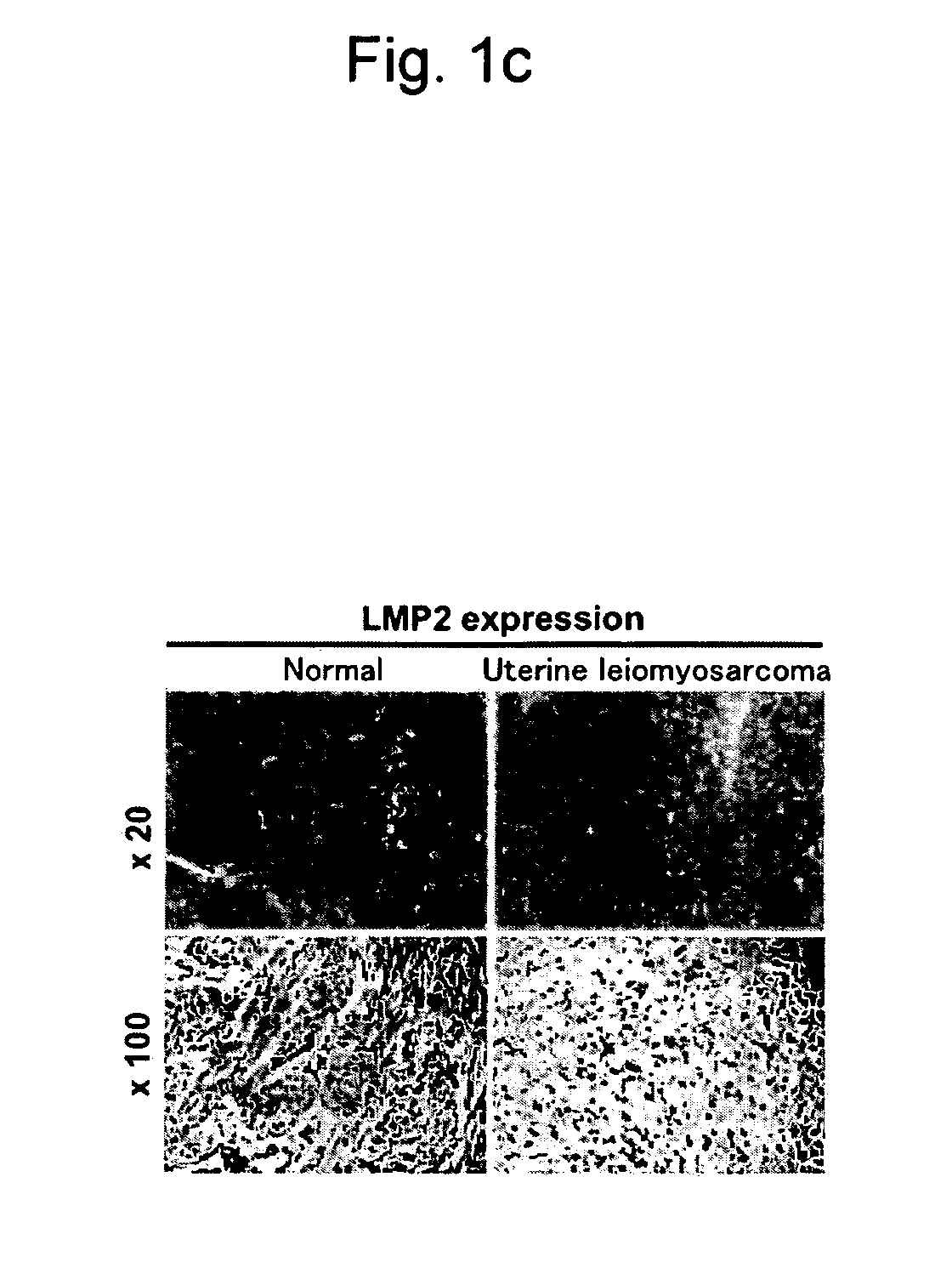
Conditions of LMP2 Expression in the Biopsy Tissue or Surgically-Removed Tissue of the Normal Human Uterine Smooth Muscle Layer, Human Uterine Leiomyoma, and Human Uterine Leiomyosarcoma via Immunohistochemical Assay
Microscopic Observation of the Human Normal Uterine Smooth Muscle Layer, Human Uterine Leiomyoma, and Human Uterine Leiomyosarcoma
[0161]Human uterine smooth muscle tissue was collected by biopsy or surgery to subject the tissue to microscopic observation.
[0162]FIG. 3 is a microscopic photograph showing the normal human uterine smooth muscle tissue, FIG. 4 is a microscopic photograph showing the human uterine leiomyoma and human uterine leiomyosarcoma, and FIG. 5 is a microscopic photograph showing the human endometrial stromal sarcoma.
[0163]Ten cases of normal uterine smooth muscle, 6 cases of uterine leiomyoma, 6 cases of endometrial sarcoma, 3 cases of uterine leiomyosarcoma (low-malignancy), and 4 cases of uterine leiomyosarcoma (high-malignancy) evaluated via the abo...
example 3
Forced Expression of LMP2 in SKN Cells
1. Changes in Cellular Morphology Upon Forced Expression of LMP2 in SKN Cells
[0169]Whether or not a lowering in LMP2 expression was directly correlated with transformation (canceration) in uterine smooth muscle cells was examined. LMP2 was forced to express via genetic recombination in the uterine leiomyosarcoma (SKN) cells in which no LMP2 expression was observed, and the configuration of SKN cells was observed.
Method
[0170]The LMP2 expression plasmid vector (2 μg) was introduced into 5×10⁵ SKN cells using FuGene6 (Roche, Indianapolis, Ind.) in accordance with the manufacturer's protocol. G418 (neomycin) was added to the culture solution 3 days after the introduction of the plasmid vector into the cell (final concentration: 200 μg / ml), and SKN cells into which the LMP2 expression plasmid vector had been introduced were exclusively selected. The cell proliferative capacity and the cell morphogenic capacity of SKN cells in which LMP2 genes had bee...
PUM
| Property | Measurement | Unit |
|---|---|---|
| thickness | aaaaa | aaaaa |
| concentration | aaaaa | aaaaa |
| doubling time | aaaaa | aaaaa |
Abstract
Description
Claims
Application Information
 - R&D
- Intellectual Property
- Life Sciences
- Materials
- Tech Scout
- Unparalleled Data Quality
- Higher Quality Content
- 60% Fewer Hallucinations
Browse by: Latest US Patents, China's latest patents, Technical Efficacy Thesaurus, Application Domain, Technology Topic, Popular Technical Reports.
© 2025 PatSnap. All rights reserved.Legal|Privacy policy|Modern Slavery Act Transparency Statement|Sitemap|About US| Contact US: help@patsnap.com