System and methods for generating a brightfield image using fluorescent images

a technology of fluorescent images and system methods, applied in the field of system and methods for generating a brightfield image using fluorescent images, can solve the problem that the diagnosis of histopathological abnormalities based on fluorescent images is not a common practi

Active Publication Date: 2011-03-31
LEICA MICROSYSTEMS CMS GMBH
View PDF9 Cites 54 Cited by
  • Summary
  • Abstract
  • Description
  • Claims
  • Application Information

AI Technical Summary

Benefits of technology

In one embodiment, a method for generating a brightfield type image, that resembles a brightfield staining protocol of a biological sample, using fluorescent images is provided for. The method comprises the steps of acquiring two or more fluorescent images of a fixed area on a biological sample, mapping said fluorescent image into a brightfield color space, and generating a bright field image.
In another embodiment an image analysis system for generating a brightfield type image, that resembles a brightfield staining protocol of a biological sample, using fluorescent images is provided. The system comprises a digital imaging device adapted to acquire two or more fluorescent images of a fixed area on a biological sample, and a processing device adapted to apply mapping parameters to transform the two or more fluorescent images into a brightfield type image.

Problems solved by technology

However, histopathological diagnosis based on fluorescent images is not a common practice because fluorescent images do not provide structural and morphological details that are essential for pathologists to diagnose.

Method used


Image

  • System and methods for generating a brightfield image using fluorescent images
  • System and methods for generating a brightfield image using fluorescent images
  • System and methods for generating a brightfield image using fluorescent images

Examples


example

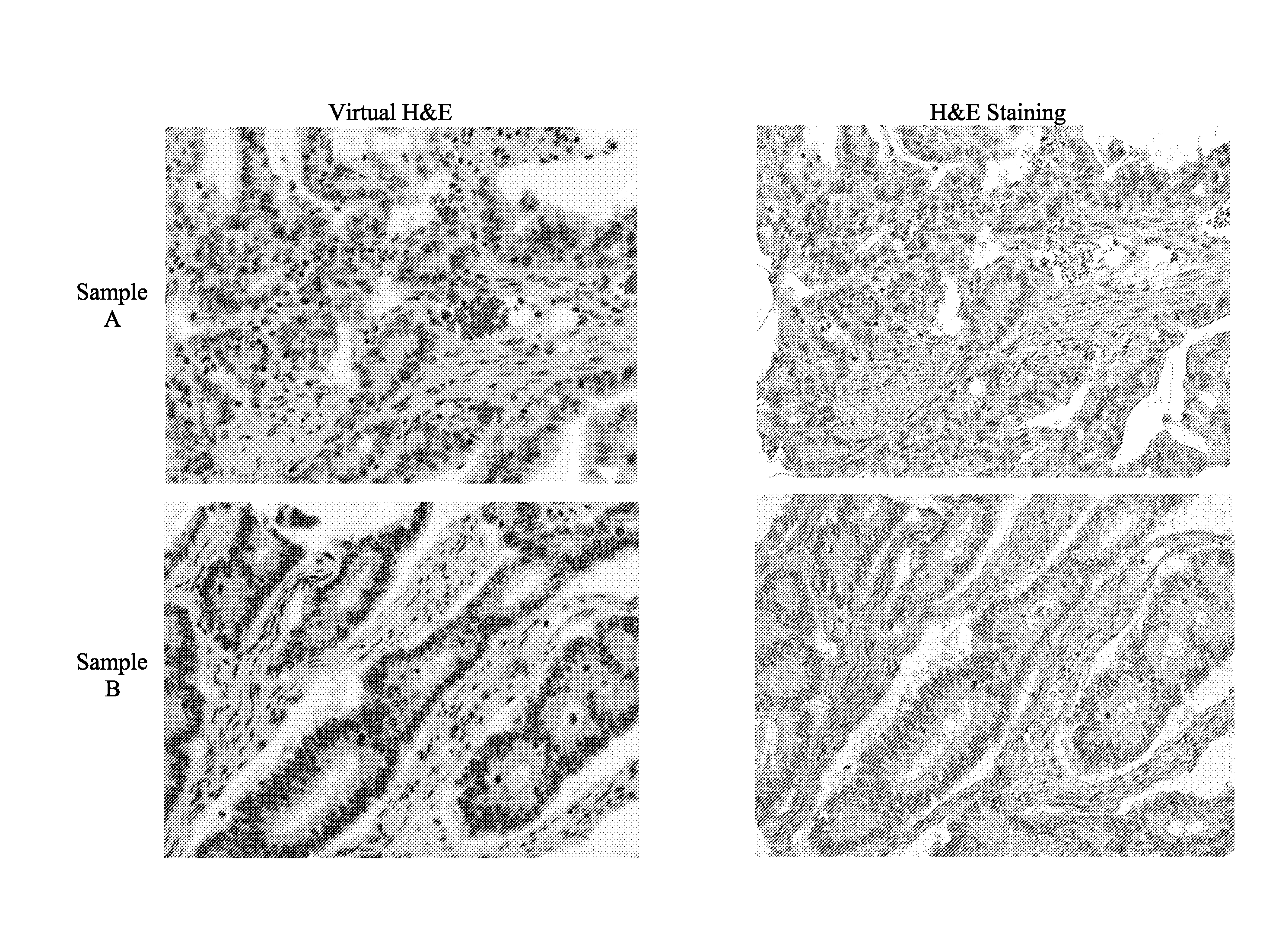
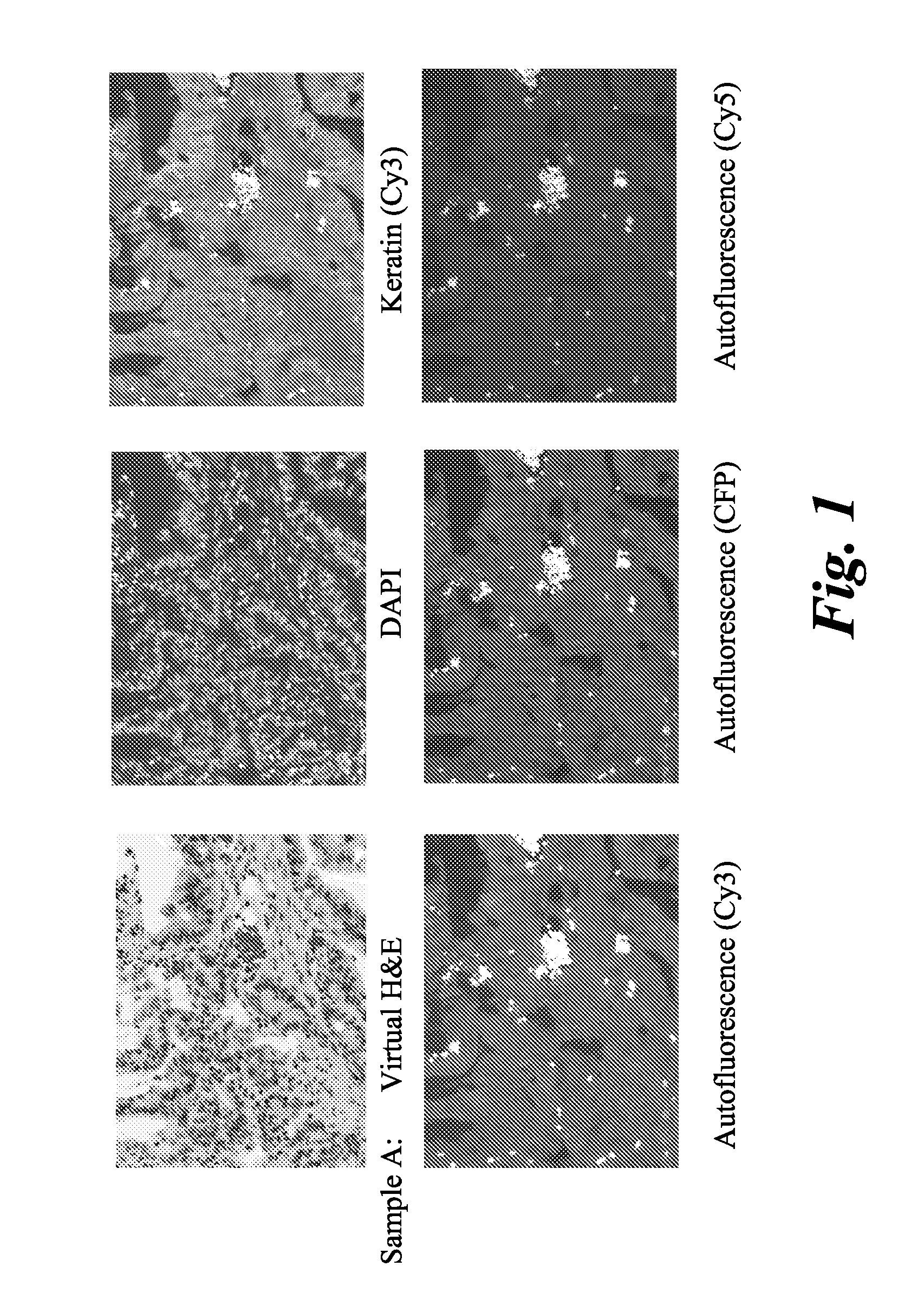
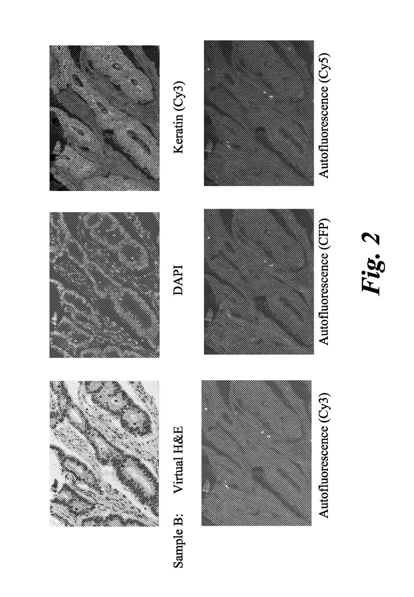
Comparison of H&E Images for Colon Tissue Samples

Adult human colon tissue samples (Biochain, T2234090) were obtained as tissue slides embedded in paraffin. Paraffin embedded slides, of adult human tissue, were subjected to an immunohistochemistry protocol to prepare them for staining. The protocol included deparaffinization, rehydration, incubation, and wash. Deparaffinization was carried by washing the slides with Histochoice (or toluene) for a period of 10 minutes and with frequent agitation. After deparaffinization, the tissue sample was rehydrated by washing the slide with ethanol solution. Washing was carried out with three different solutions of ethanol with decreasing concentrations. The concentrations of ethanol used were 90 volume %, 70 volume %, and 50 volume %. The slide was then washed with a phosphate buffer saline (PBS, pH 7.4). Membrane permeabilization of the tissue was carried out by washing the slide with 0.1 weight percent solution of Triton TX-100. Citrate buffer...


PUM


Abstract

A method for generating a brightfield type image, which resembles a brightfield staining protocol of a biological sample, using fluorescent images is provided. The steps comprise acquiring two or more fluorescent images of a fixed area on a biological sample, mapping said fluorescent image into a brightfield color space, and generating a bright field image. Also provided is an image analysis system for generating a brightfield type image of a biological sample using fluorescent images.

Description

BACKGROUNDThe invention relates generally to a method to map a set of biomarker images acquired by a fluorescent microscope into a new color space where the mapped image intensity values represent a brightfield modality.In traditional histological staining with Hematoxylin & Eosin (H&E), the basophilic dye Hematoxylin (H) is used to stain the cell nuclei blue, and the acidophilic dye Eosin (E) is used as a counter-stain to stain cytoplasm, connective tissue (collagen), muscle fibers, connective tissue, and red blood cells. Eosin interacts with different cellular components in the tissue producing different shades of pink color based on charge properties of the molecules to which eosin is binding. These cellular components can be alternatively labeled using molecular markers (dyes and antibodies) with fluorescent dyes. For example, cell nuclei can be stained with DAPI (a fluorescent dye that binds DNA specifically) while other regions in the tissue can be labeled immunofluorescently ...

Claims


Application Information

Patent Timeline
Patent Type & Authority Applications(United States)
IPC IPC(8): H04N7/18G06K9/00
CPCG01N21/6458G06T5/50G06T2207/30024G06T2207/10064G06T2207/20221G06T2207/10056G01N21/6428G01N21/6486G01N2021/6423G06V20/693G01N21/64G01N33/483G06T7/00
Inventor CAN, ALIBELLO, MUSODIQ O.GERDES, MICHAEL JOHNLI, QING
Owner LEICA MICROSYSTEMS CMS GMBH
Why Eureka
Try Eureka