Radiation imaging method with individual signal resolution

a radiation imaging and signal resolution technology, applied in the field of imaging methods and equipment, can solve the problems of poor image quality and general count, and achieve the effects of reducing radiation exposure, spatial resolution, and reducing tracer dose or concentration

Active Publication Date: 2011-06-23
SOUTHERN INNOVATION INT
View PDF26 Cites 19 Cited by
  • Summary
  • Abstract
  • Description
  • Claims
  • Application Information

AI Technical Summary

Benefits of technology

[0012]whereby acceptable subject to detector distance is reduced or increased, spatial resolution is improved, tracer dose or concentration is reduced, subject radiation exposure is reduced and / or scanning time is reduced.
[0088]whereby acceptable subject to detector distance is reduced or increased, spatial resolution is improved, tracer dose or concentration is reduced, subject radiation exposure is reduced and / or scanning time is reduced.

Problems solved by technology

Image quality is, therefore, generally count poor.

Method used


Image

  • Radiation imaging method with individual signal resolution
  • Radiation imaging method with individual signal resolution
  • Radiation imaging method with individual signal resolution

Examples


example

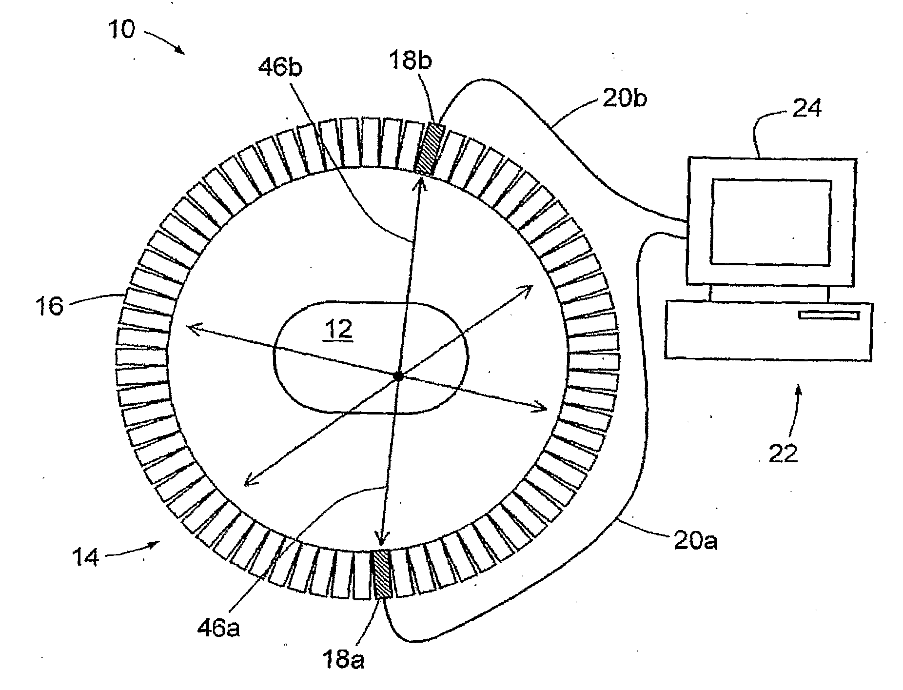
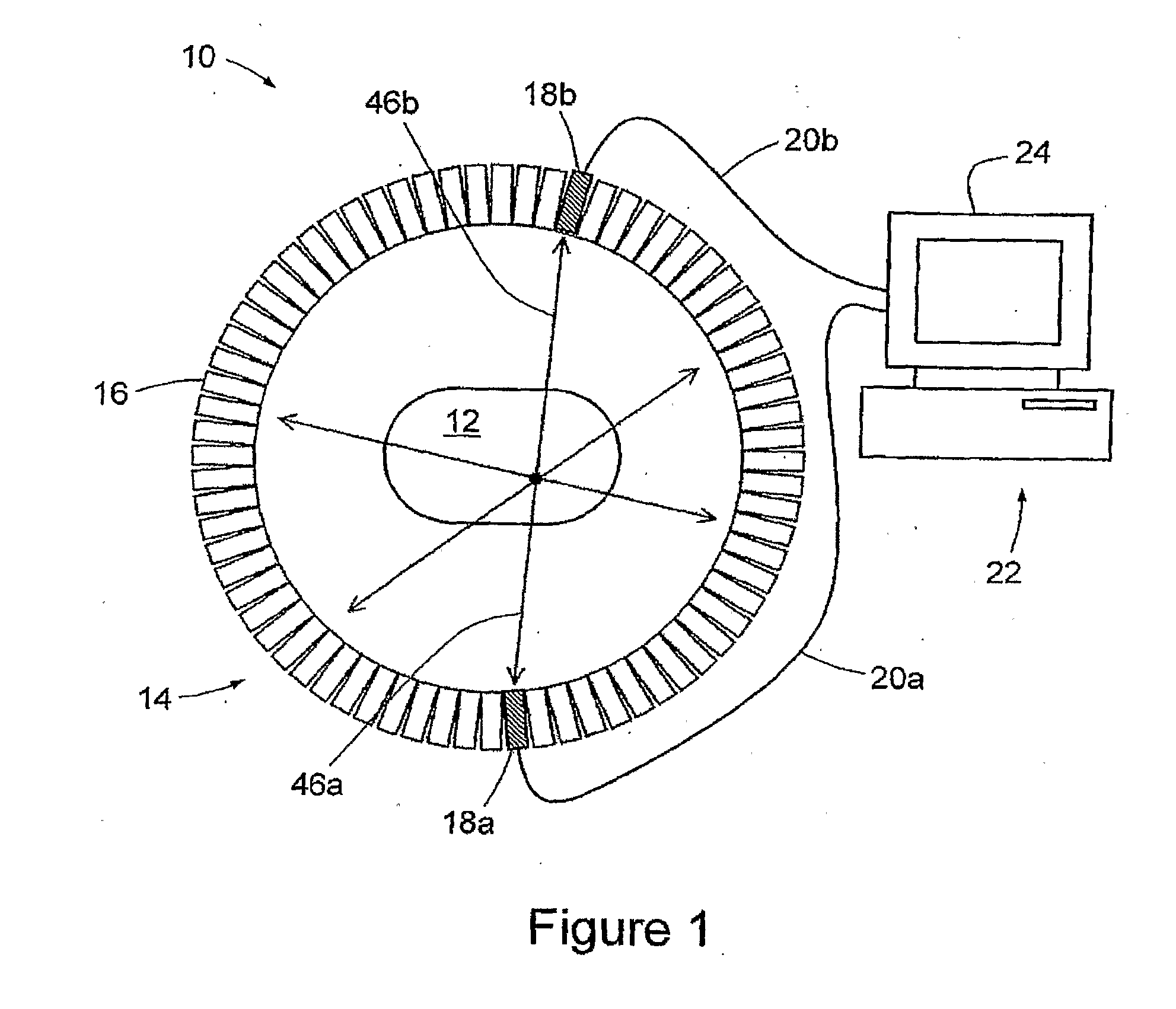
[0194]X-ray computed tomography is a medical imaging technique that uses a computer to stitch together multiple 2D images (projections) into a 3D image. FIG. 21 is a schematic view of a CT machine 304 according to an embodiment of the present invention. CT machine 304 includes an X-ray source in the form of an X-ray generator 306, to produce a fan beam of X-rays 308, and X-ray detectors 310 positioned on the opposite side of the machine from source 306 for detecting X-rays from X-ray generator 306 that have interacted with a subject 312 (shown as a recumbent patient). In use, projections are generated by using X-ray generator 306 to produce X-rays 308, and to detect X-rays with X-ray detectors 310. Both X-ray generator 306 and detectors 310 rotate around the subject, while maintaining their relative positions.

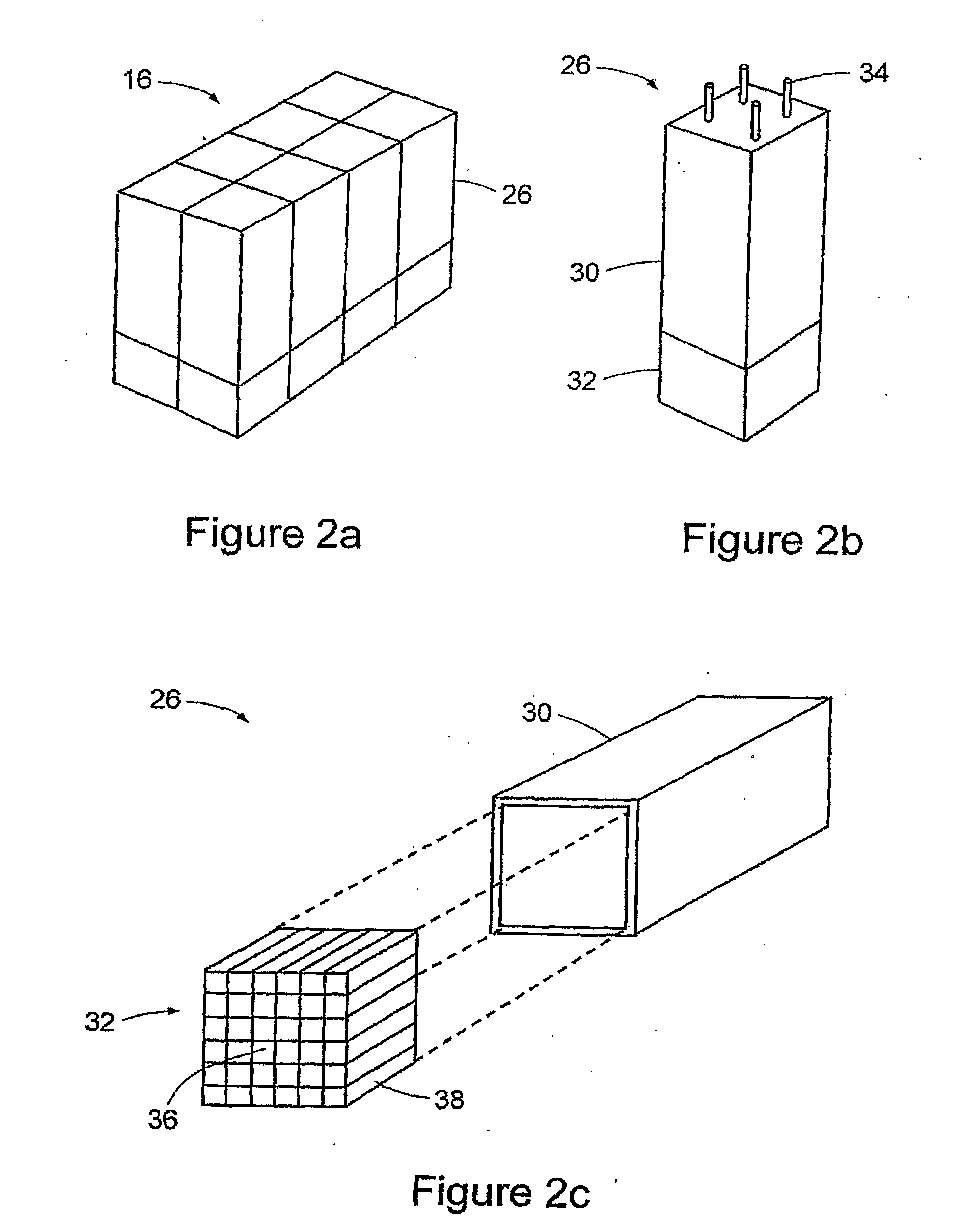
[0195]FIG. 22a is a schematic view of an exemplary detector 314 of detectors 310 of CT machine 304. There may be multiple detectors used in one CT machine in order to capture m...


PUM


Abstract

An imaging method and apparatus, the method comprising collecting detector output data from a radiation detector positioned near a subject provided with a radio-active tracer, and resolving individual signals in the detector output data by (i) determining a signal form of signals present in the data, (ii) making parameter estimates of one or more parameters of the signals, wherein the one or more parameters comprise at least a signal temporal position, and (iii) determining the energy of each of the signals from at least the signal form and the parameter estimates. The acceptable subject to detector distance is reduced or increased, spatial resolution is improved, tracer dose or concentration is reduced, subject radiation exposure is reduced and / or scanning time is reduced.

Description

RELATED APPLICATION[0001]This application is based on and claims the benefit of the filing date of U.S. application No. 61 / 041,144 filed 31 Mar. 2008 and of U.S. application No. 61 / 138,879 filed 18 Dec. 2008, the contents of which as filed are incorporated herein by reference in their entirety.FIELD OF THE INVENTION[0002]The present invention relates generally to an imaging method and apparatus, of particular but by no means exclusive application in medical imaging techniques such as Single Photon Emission Computed Tomography (SPECT), Positron Emission Tomography (PET) and gamma-camera imaging.BACKGROUND OF THE INVENTION[0003]Molecular Imaging is emerging as a powerful and sensitive technique for functional imaging of the biological functions of the human body. In particular Single Photon Emission Computed Tomography (SPECT) and Positron Emission Tomography (PET) are two of the most effective technologies for the detection and staging of cancer.[0004]In these techniques, tracers com...

Claims


Application Information

Patent Timeline
Patent Type & Authority Applications(United States)
IPC IPC(8): G01T1/20G01T1/16
CPCA61B6/037G01T1/171G01T1/1644
Inventor SCOULLAR, PAUL ANDREW BASILEVANS, ROBIN JOHNMCLEAN, CHRISTOPHER CHARLES
Owner SOUTHERN INNOVATION INT
Why Eureka
Try Eureka