Methods and compositions for nuclear staining

a technology of compositions and nuclear staining, which is applied in the field of methods and compositions for nuclear staining, can solve the problems of limited stability in solution, incompatibility of nuclear fast red with all the above mentioned staining procedures, etc., and achieve the effect of superior tissue architecture and cellular detail

Inactive Publication Date: 2011-09-22
UNIVERSITY OF ROCHESTER
View PDF8 Cites 10 Cited by
  • Summary
  • Abstract
  • Description
  • Claims
  • Application Information

AI Technical Summary

Benefits of technology

[0009]As described herein, the composition of the present invention offers many advantages over currently available nuclear dye solutions such as nuclear fast red. Firstly, the composition of the present invention does not precipitate in solution and has a shelf-life of at least one year. In addition, nuclear staining with the composition of the present invention achieves brighter, more brilliant nuclear staining showing superior tissue architecture and cellular detail within seconds of exposure. Finally, unlike conventional dyes that can weaken within weeks of staining, the nuclear staining composition of the present invention is extremely lightfast, with no significant fading observed over time.

Problems solved by technology

While nuclear fast red is most commonly used in methods requiring a red nuclear counterstain, this dye involves incubations periods of 5-10 minutes, and often fades within just weeks of staining.
In addition, nuclear fast red is not compatible with all of the above noted staining procedures and has limited stability in solution.

Method used


Image

  • Methods and compositions for nuclear staining
  • Methods and compositions for nuclear staining
  • Methods and compositions for nuclear staining

Examples


example 1

Preparation of 0.1% Pararosanilin Solution

[0033]The 0.1% pararosanilin solution of the present invention is prepared by combining 0.1 gram pararosanilin (C.I. 52000) with 100 mL 0.6% lactic acid solution and 0.1 mL of 25% polysorbate 20. The pH of this solution is approximately 2.50.

example 3

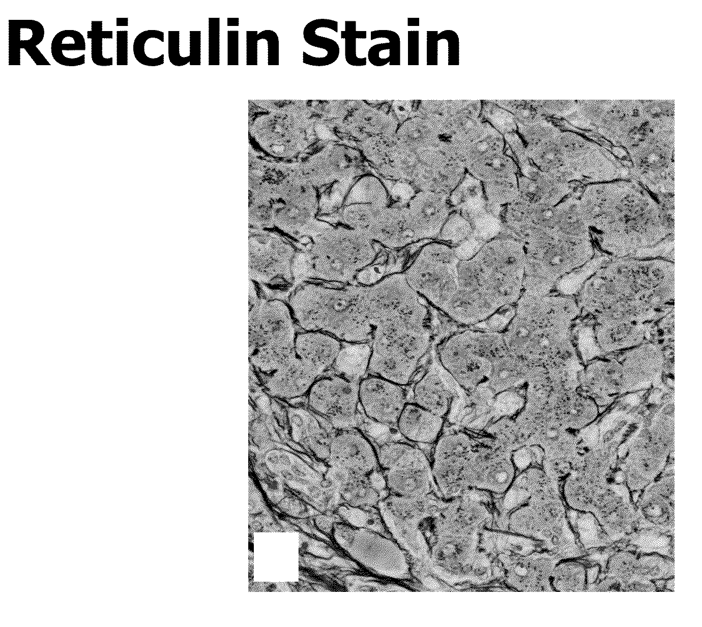
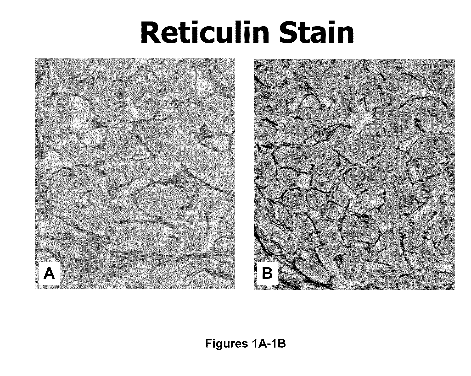
Modified Gomori's Method for Reticulin Staining with Nuclear Counterstain in Liver Tissue Sections

[0035]Liver tissue samples were fixed in 10% buffered neutral formalin and embedded in paraffin. Paraffin sections were cut at 5 μm. For reticulum staining, sections were deparaffinized, hydrated, and then oxidized using acidified potassium permanganate (0.3 gm potassium permanganate, 100 mL distilled water, and 0.2 mL sulfuric acid) for three minutes. Sections were rinsed in distilled water and reduced with 1% potassium metabisulfite for 1 minute. Sections were rinsed with running tap water for three minutes and then rinsed with four changes of distilled water prior to incubating with Ammoniacal silver solution for two minutes. Tissue sections were then rinsed three times and reduced in 10% formalin for one minute. Following the incubation in formalin, the sections were washed with running tap water for one minute and then rinsed with two changes of distilled water. Next, sections were...

example 4

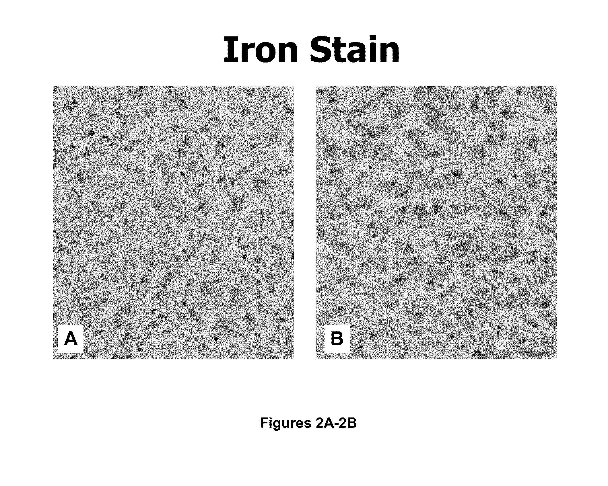
Perl's Method for Ferric Iron Staining with Nuclear Counterstain in Liver Tissue Sections

[0038]Liver tissue was fixed in 10% buffered neutral formalin, embedded in paraffin, and cut at 5 μm for staining. Slides containing the liver tissue sections were deparaffinized and hydrated in distilled water. Slides were then placed in hydrochloric acid-potassium ferrocyanide solution (20 mL 2% hydrochloric acid and 20 mL 1% potassium ferrocyanide) for 30 minutes at room temperature. Following this incubation, the slides were rinsed in five changes of distilled water and counterstained with 0.1% nuclear fast red solution of Comparative Example 2 for 5 minutes or the 0.1% pararosaniline solution of Example 1 for 10 seconds. Sections were rinsed, dehydrated, cleared in xylene, and mounted with synthetic resin.

[0039]FIGS. 2A and 2B show a comparison of the ferric iron staining in liver tissue with nuclear fast red counterstain (FIG. 2A) and 0.1% pararosaniline nuclear counterstain (“strong fast ...


PUM

PropertyMeasurementUnit
exposure timeaaaaaaaaaa
pHaaaaaaaaaa

Abstract

The present invention relates to compositions, methods, and kits suitable for detecting nucleic acids in a biological sample. The nuclear staining composition of the present invention contains a pH buffering reagent, a solubilizing reagent, a basic dye, and an aqueous medium. The composition can be used alone to detect nucleic acids in a biological sample or in combination with other histological dyes for nuclear counterstaining.

Description

[0001]This application claims the benefit of U.S. Provisional Patent Application Ser. No. 61 / 315,483, filed Mar. 19, 2010, which is hereby incorporated by reference in its entirety.FIELD OF THE INVENTION[0002]The present invention relates to compositions and methods suitable for detecting nuclear elements in a biological sample.BACKGROUND OF THE INVENTION[0003]Histochemical procedures performed on surgical, autopsy, and biopsy tissue and cell samples for diagnostic and research purposes generally involve the use of a nuclear counterstain to delineate tissue architecture and cellular detail. The nuclear counterstain is usually a dark color to contrast a lighter dye used to label the cytoplasmic or extracellular structures of interest.[0004]A nuclear counterstain is the desired stain for a number of histological staining procedures including, for example, Gomori's reticulum, Pearl's ferric iron, Alcian blue for acidic mucins, Jones basement membrane, Churukian's ammonical silver for m...

Claims


Application Information

Patent Timeline
Patent Type & Authority Applications(United States)
IPC IPC(8): C12Q1/68
CPCG01N33/5308G01N33/52
Inventor CHURUKIAN, CHARLES J.
Owner UNIVERSITY OF ROCHESTER
Why Eureka
Try Eureka