Transcatheter Heart Valve Delivery System With Reduced Area Moment of Inertia

a delivery system and transcatheter technology, applied in the field of systems and methods for percutaneous implantation of heart valve prosthesis, can solve the problems of inability to achieve mixed results, inability to achieve significant patient trauma and discomfort, and inability to achieve significant clinical benefits, etc., to achieve sufficient conformability, reduce the area moment of inertia, and the radial stiffness of the capsule distal segment is greater.

Active Publication Date: 2011-10-13
MEDTRONIC INC
View PDF8 Cites 94 Cited by
  • Summary
  • Abstract
  • Description
  • Claims
  • Application Information

AI Technical Summary

Benefits of technology

[0009]Some aspects in accordance with principles of the present disclosure relate to a device for repairing a heart valve of a patient. The device includes a prosthetic heart valve and a delivery system. The prosthetic heart valve has a stent frame and a valve structure. The valve structure is attached to the stent frame and forms at least two valve leaflets. Further, the prosthetic heart valve is radially self-expandable from a compressed arrangement to a natural arrangement, with the stent frame defining an axial length. The delivery system includes an inner shaft assembly and a delivery sheath. The inner shaft assembly includes an intermediate portion forming a coupling structure. The delivery sheath assembly defines a lumen sized to slidably receive the inner shaft assembly, and includes a shaft and a capsule. The capsule extends distally from the shaft and includes a distal segment and a proximal segment. An outer diameter of the capsule distal segment is greater than an outer diameter of the capsule proximal segment. Further, an axial length of the distal segment is less than the axial length of the stent frame. With this in mind, the device is configured to provide a loaded state in which the prosthetic heart valve engages the coupling structure and is compressively retained in the compressed arrangement within the capsule. In some embodiments, an inner diameter of the capsule distal segment is greater than an inner diameter of the capsule proximal segment. In other embodiments, a radial stiffness of the capsule distal segment is greater than a radial stiffness of the capsule proximal segment. In yet other embodiments, an area moment of inertia of the distal segment is greater than area moment of inertia of the proximal segment. In yet other embodiments, in the loaded state, an inflow side of the prosthetic heart valve is crimped within the distal segment of the capsule, and an outflow side of the prosthetic heart valve is crimped within the proximal segment. With these and other constructions, the capsule provides requisite resistance to radial expansion of the prosthetic heart valve, yet exhibits sufficient conformability for traversing the tortuous vasculature of the patient, for example the various curvatures of the aortic arch. For example, by providing the capsule with a lower area moment of inertia (as compared to the outer sheath capsule of a conventional self-expanding stented prosthetic heart valve delivery system), the devices of the present disclosure greatly reduce the likelihood of the capsule becoming kinked when tracking around the aortic arch. This feature, in turn, facilitates easier recapturing of the prosthetic heart valve with the capsule if desired.

Problems solved by technology

Traditional open surgery inflicts significant patient trauma and discomfort, and exposes the patient to a number of potential risks, such as infection, stroke, renal failure, and adverse effects associated with the use of the heart-lung bypass machine, for example.
Although transcatheter techniques have attained widespread acceptance with respect to the delivery of conventional stents to restore vessel patency, only mixed results have been realized with percutaneous delivery of a relatively more complex prosthetic heart valve.
If the prosthesis is incorrectly positioned relative to the native annulus, serious complications can result as the deployed device can leak and may even dislodge from the implantation site.
While imaging technology can be employed as part of the implantation procedure to assist a clinician in better evaluating a location of the transcatheter prosthetic heart valve immediately prior to full deployment, in many instances this evaluation alone is insufficient.
While, in theory, the recapturing of a partially deployed stented prosthetic heart valve is straight forward, in actual practice, the constraints presented by the implantation access path and the stented prosthetic heart valve itself render the procedure exceedingly difficult.
While the relatively rigid, relatively large delivery sheath capsules are viable for accessing the native heart valve via the aortic arch (or other tortuous vasculature), the so-configured delivery sheath may undesirably buckle or “kink”, especially when traversing the various curvatures of the aortic arch.
Once kinked, it is virtually impossible for the delivery sheath capsule to be advanced over a partially-deployed prosthesis as is otherwise necessary for recapture.
Simply stated, due to the relatively long stiff section of the conventional delivery sheath, successful delivery of a prosthetic heart valve through the tortuous vasculature, such as required for retrograde delivery of a prosthetic aortic heart valve, as well as recapturing a partially deployed prosthetic heart valve, has proven to be difficult.

Method used


Image

  • Transcatheter Heart Valve Delivery System With Reduced Area Moment of Inertia
  • Transcatheter Heart Valve Delivery System With Reduced Area Moment of Inertia
  • Transcatheter Heart Valve Delivery System With Reduced Area Moment of Inertia

Examples


Embodiment Construction

[0024]As referred to herein, the prosthetic heart valve as used in accordance with the various devices and methods may include a wide variety of different configurations, such as a bioprosthetic heart valve having tissue leaflets or a synthetic heart valve having a polymeric, metallic, or tissue-engineered leaflets, and can be specifically configured for replacing any heart valve. Thus, the prosthetic heart valve useful with the devices and methods of the present disclosure can be generally used for replacement of a native aortic, mitral, pulmonic, or tricuspid valves, for use as a venous valve, or to replace a failed bioprosthesis, such as in the area of an aortic valve or mitral valve, for example.

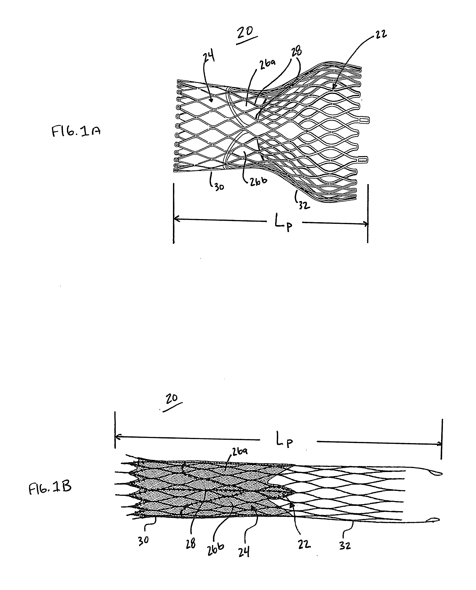
[0025]In general terms, the prosthetic heart valves of the present disclosure include a stent or stent frame maintaining a valve structure (tissue or synthetic), with the stent having a normal, expanded arrangement or state and collapsible to a compressed arrangement for loading within t...


PUM


Abstract

A device for percutaneously repairing a heart valve of a patient including a self-expanding, stented prosthetic heart valve and a delivery system. The delivery system includes delivery sheath slidably receiving an inner shaft forming a coupling structure. A capsule of the delivery sheath includes a distal segment and a proximal segment. An outer diameter of the distal segment is greater than that of the proximal segment. An area moment of inertia of the distal segment can be greater than an area moment of inertia of the proximal segment. Regardless, an axial length of the distal segment is less than the axial length of the prosthesis. In a loaded state, the prosthesis engages the coupling structure and is compressively retained within the capsule. The capsule is unlikely to kink when traversing the patient's vasculature, such as when tracking around the aortic arch, promoting recapturing of the prosthesis.

Description

BACKGROUND[0001]The present disclosure relates to systems and methods for percutaneous implantation of a heart valve prosthesis. More particular, it relates to delivery systems and methods for transcatheter implantation of a self-expanding, stented prosthetic heart valve.[0002]Diseased or otherwise deficient heart valves can be repaired or replaced with an implanted prosthetic heart valve. Conventionally, heart valve replacement surgery is an open-heart procedure conducted under general anesthesia, during which the heart is stopped and blood flow is controlled by a heart-lung bypass machine. Traditional open surgery inflicts significant patient trauma and discomfort, and exposes the patient to a number of potential risks, such as infection, stroke, renal failure, and adverse effects associated with the use of the heart-lung bypass machine, for example.[0003]Due to the drawbacks of open-heart surgical procedures, there has been an increased interest in minimally invasive and percutan...

Claims


Application Information

Patent Timeline
Patent Type & Authority Applications(United States)
IPC IPC(8): A61F2/24
CPCA61F2/2418A61F2/2436A61F2250/0039A61F2250/0018
Inventor TRAN, DONWIEMEYER, NATHANDESHMUKH, SUSHEELMENDOZA, LEONALRUST, MATTHEW
Owner MEDTRONIC INC
Why Eureka
Try Eureka