Magnetic resonance imaging water-fat separation method

a magnetic resonance imaging and water-fat separation technology, applied in the field of magnetic resonance imaging, can solve the problems of increasing image calculation complexity and time, and shortening the entire imaging time, so as to improve the efficiency of the mri device, reduce the mri scan time, and shorten the entire imaging time

Active Publication Date: 2011-11-03
SIEMENS HEALTHCARE GMBH
View PDF13 Cites 31 Cited by
  • Summary
  • Abstract
  • Description
  • Claims
  • Application Information

AI Technical Summary

Benefits of technology

From the above technical solution it can be seen that the present invention reconstructs the in-phase and out-of-phase images by using the penalty function regularized iterative method, which eliminates the need for k-space raw measurement data with 100% sampling rate, so as to reduce the MRI scan time, shorten the entire imaging time, and improve the efficiency of the MRI device. Additionally, in the present invention the process of reconstructing the in-phase image is independent of constructing the out-of-phase image, thus the present invention can rebuild the in-phase image and out-of-phase image in parallel, which further reduces the imaging time and improves the imaging efficiency of the magnetic resonance imaging device.

Problems solved by technology

In magnetic resonance imaging (MRI), the body's molecular environment for hydrogen protons in fat tissue is different from that for hydrogen protons in other tissues, which results in a difference in the resonance frequency of the protons.
The single-point Dixon method, two-point Dixon method and three-point and multiple-point Dixon methods are easy and timesaving, but they are sensitive to motion artifacts, and the spin echo sequence is also sensitive to motion artifacts, so there are usually motion artifacts in the images obtained by the Dixon method based on the Cartesian trajectory sampling.
The advantage of this kind of methods is that the motion introduces fuzziness rather than artifacts into the reconstructed image, which has little impact on identifying the objects in the image, but using the radial or spiral trajectory sampling usually increases the image calculation complexity and takes much more time.
The radial or spiral trajectory sampling methods convert the motion artifact into fuzziness in the reconstructed image, but the calculation is complex and it takes much more time.
However, the gridding reconstructing method needs a high sampling rate (usually 100% sampling rate) to eliminate the strip artifacts, so it needs a longer scan time to acquire k-space raw measurement data, which causes the entire imaging to take longer and reduces the efficiency of the MRI device.

Method used


Image

  • Magnetic resonance imaging water-fat separation method
  • Magnetic resonance imaging water-fat separation method
  • Magnetic resonance imaging water-fat separation method

Examples


Embodiment Construction

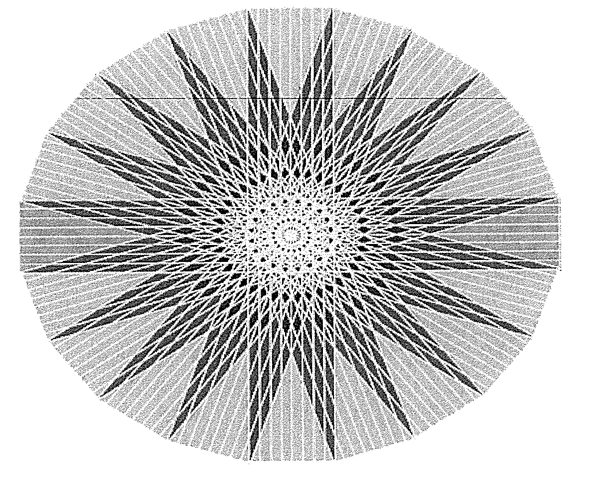
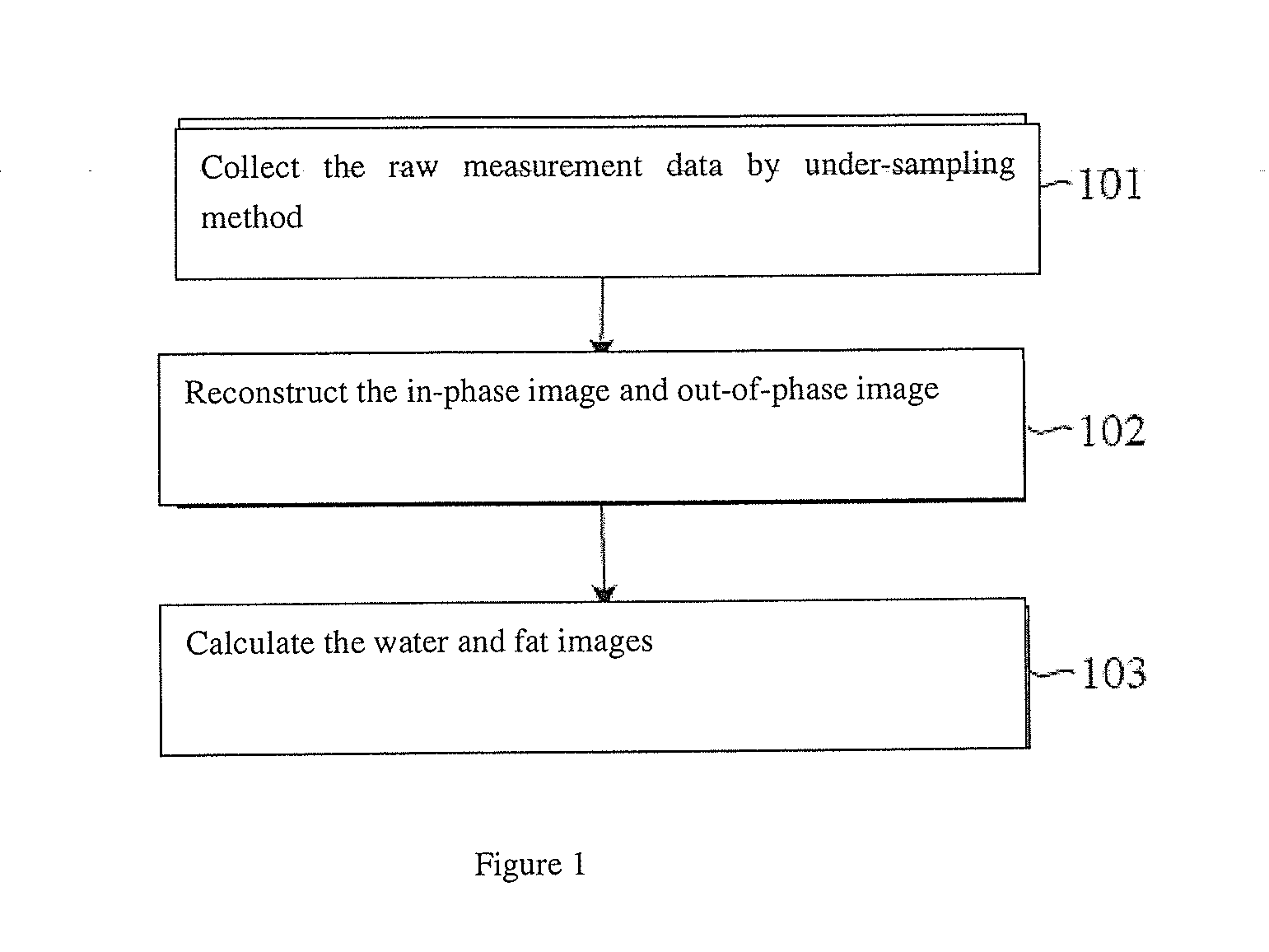
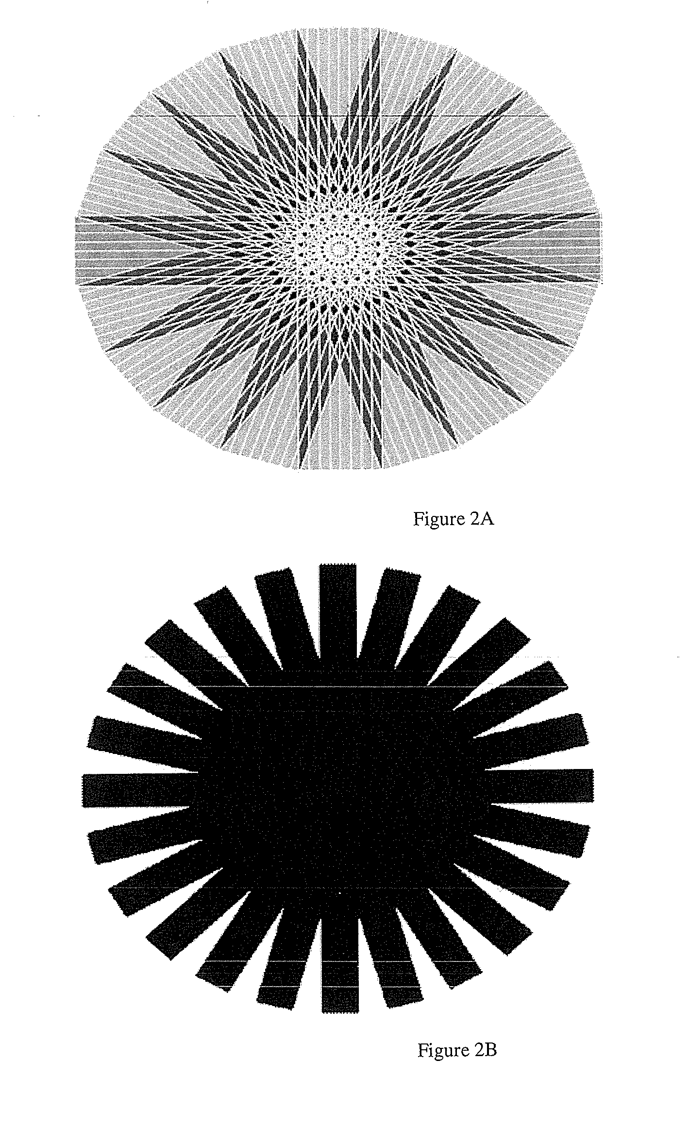
FIG. 1 is a schematic diagram illustrating the flow of one embodiment according to the present invention. In this embodiment, the under-sampling BLADE trajectory is used to acquire k-space raw measurement data, and the water and fat images are calculated based on the three-point Dixon method. However, the present invention is not limited to this technique, and other k-space trajectories can be used to acquire the raw measurement data, and the water and fat images can be calculated based on other algorithms (e.g. two-point Dixon method, etc.).

Referring to FIG. 1, the MRI water-fat separation method according to the embodiment of the present invention includes the following steps:

Step 101, acquiring the raw measurement data of one in-phase image and two out-of-phase images.

In order to understand the present invention easily, the solution of the present invention will be described in detail by using the example of using BLADE trajectory to acquire k-space raw measurement data. It shoul...


PUM


Abstract

A magnetic resonance imaging (MRI) water-fat separation method includes acquiring in-phase image raw measurement data and out-of-phase image raw measurement data with an MRI device, reconstructing an in-phase image and an out-of-phase image according to a system matrix and the raw measurement data using the penalty function regularized iterative reconstruction method, and calculating water and fat images according to the in-phase image and the out-of-phase image. The use of the penalty function regularized iterative method eliminates the need for k-space raw measurement data with a 100% sampling rate, thereby reducing the MRI scan time, shortening the entire imaging time, and improving the efficiency of the MRI device.

Description

BACKGROUND OF THE INVENTION1. Field of the InventionThe present invention relates to the magnetic resonance imaging (MRI) technical field, and more particularly to an MRI water-fat separation method, wherein k-space raw measurement data are acquired based on the under-sampling method.2. Description of the Prior ArtIn magnetic resonance imaging (MRI), the body's molecular environment for hydrogen protons in fat tissue is different from that for hydrogen protons in other tissues, which results in a difference in the resonance frequency of the protons. The relaxation time of the hydrogen protons in fat tissue and those in other tissues is also different when they are excited by radio frequency pulses at the same time. When signals are collected at different echo times, the fat tissue and non-fat tissues show different phases and signal strengths.The Dixon method is known for use to generate pure water proton images in magnetic resonance imaging, and its basic principle is: acquiring th...

Claims


Application Information

Patent Timeline
Patent Type & Authority Applications(United States)
IPC IPC(8): G01R33/44
CPCA61B5/055G01R33/4828G01R33/4824
Inventor HE, QIANGWENG, DE HE
Owner SIEMENS HEALTHCARE GMBH
Why Eureka
Try Eureka