Method and Apparatus to Optimize Injected Dose and Scan Time in SPECT Imaging

a spect imaging and dose optimization technology, applied in the field of medical imaging systems and methods, can solve the problems of high malpractice insurance costs, hospitals and medical fields have to find a balance, and low cost medical services cannot be provided, so as to reduce radiation dose to patients, improve quality, and simplify steps

Active Publication Date: 2012-01-26
SIEMENS MEDICAL SOLUTIONS USA INC
View PDF2 Cites 6 Cited by
  • Summary
  • Abstract
  • Description
  • Claims
  • Application Information

AI Technical Summary

Benefits of technology

[0014]According to one embodiment of the present invention, an apparatus and method is provided for simplifying the steps in a clinical protocol and uses a minimum number of parameters but still provides high quality images.
[0015]According to another embodiment of the present invention, an apparatus and method is provided for reducing radiation dose to a patient.

Problems solved by technology

With the high cost of malpractice insurance, hospitals and the medical field have to find a balance.
Low cost medical service cannot be provided if it puts a patient at risk because it will in turn put the hospital or medical provider at risk for a law suit.
The approach provided is also complicated because it has so many options for a practitioner.
It is easy to get overwhelmed.

Method used


Image

  • Method and Apparatus to Optimize Injected Dose and Scan Time in SPECT Imaging
  • Method and Apparatus to Optimize Injected Dose and Scan Time in SPECT Imaging
  • Method and Apparatus to Optimize Injected Dose and Scan Time in SPECT Imaging

Examples


Embodiment Construction

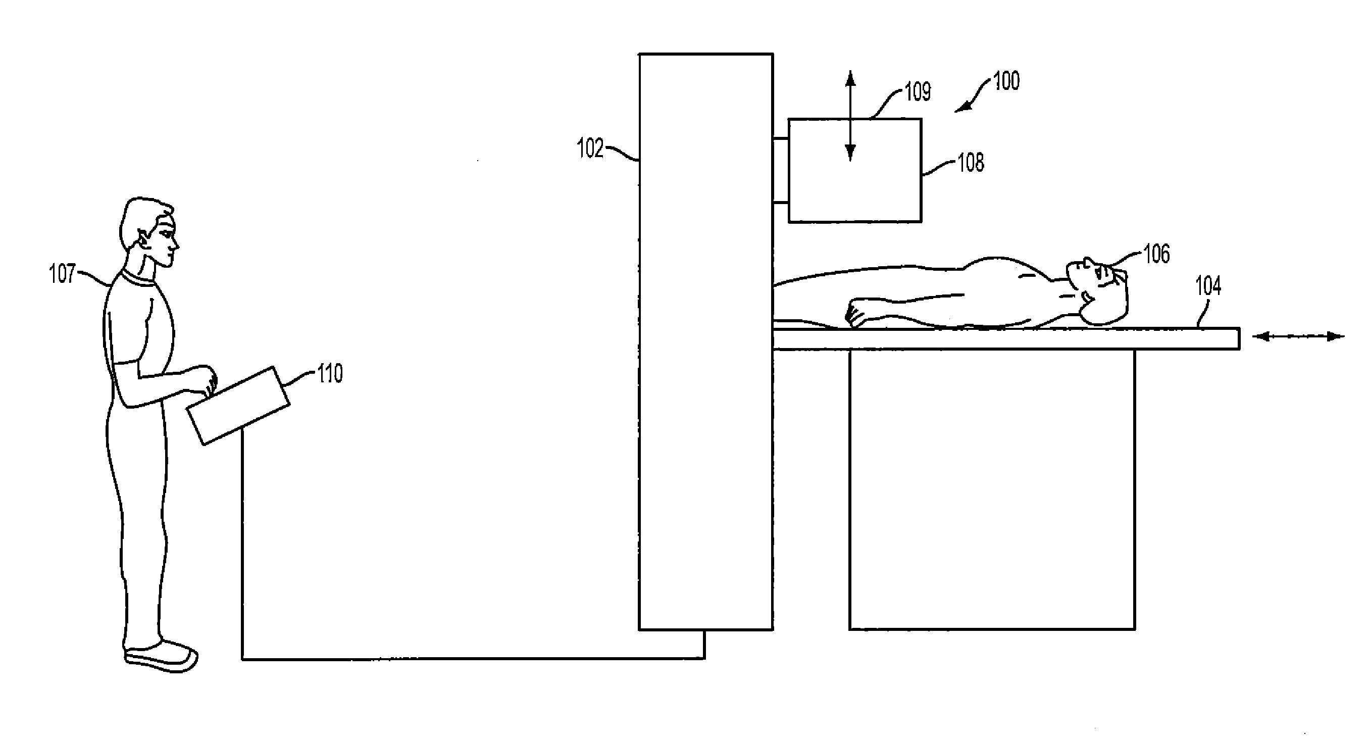
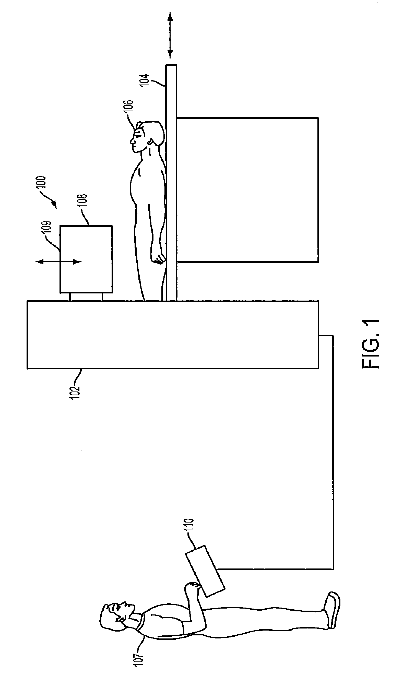
[0025]FIG. 1 depicts components of a SPECT system 100 (i.e., having a gamma or scintillation camera) which includes a gantry 102 supporting one or more detectors 108 enclosed within a metal housing and movably supported proximate a patient 106 located on a patient support (e.g., pallet or table) 104. The detectors are proximate collimators 109. The collimators a parallel beam, fan beam, multifocal collimator and the like. Typically, the positions of the detectors 108 can be changed to a variety of orientations to obtain images of a patient's body from various angles and locations along the patient's body. In many instances, a data acquisition console 110 (e.g., with a user interface and / or display) is located proximate a patient during use for a technologist 107 to manipulate during data acquisition. In addition to the data acquisition console 110, images are often “reconstructed” or developed from the acquired image data (“projection data”) via a processing computer system that is ...


PUM


Abstract

An apparatus and method are provided for optimizing an amount of radiation dose and acquisition time in cardiac Single Photon Emission Computed Tomography (SPECT) imaging. The apparatus and method include providing an organ, acquiring images of the organ at projected views. Then a projected view that projects the organ as an annulus is selected; a region of interest (ROI) is also selected in the projected view, wherein the ROI is in a lateral wall of the organ. An average count in the ROI is determined; and an image quality of a reconstructed image based on the average count is predicted.

Description

CROSS-REFERENCE TO RELATED APPLICATIONS AND CLAIM FOR PRIORITY[0001]This application claims priority under 35 U.S.C. §119(e) from copending U.S. Provisional Patent Application Ser. No. 61 / 366,257 filed Jul. 21, 2010, the entire contents of which are incorporated herein by reference.BACKGROUND OF THE INVENTION[0002]1. Field of the Invention[0003]This invention generally pertains to systems and methods for performing medical imaging. More particularly, the present invention is directed to an apparatus and method for optimizing an injected dose and scan time in SPECT imaging.[0004]2. Description of the Related Art[0005]The medical imaging field is improving. For example, image quality has greatly improved from a few years ago. In some cases, the changes are dramatic. Images are very life like. This has come about due to improvements in medical equipment design such as collimators and in software such as the reconstruction software that reproduces the images.[0006]However, external fact...

Claims


Application Information

Patent Timeline
Patent Type & Authority Applications(United States)
IPC IPC(8): G01T1/164
CPCA61B6/037A61B6/545A61B6/542A61B6/481
Inventor VIJA, ALEXANDER HANS
Owner SIEMENS MEDICAL SOLUTIONS USA INC
Why Eureka
Try Eureka