
Brain Tumor Stem Cell Differentiation Promoter, and Therapeutic Agent for Brain Tumor

Inactive Publication Date: 2012-02-16
THE UNIV OF TOKYO
View PDF1 Cites 1 Cited by
  • Summary
  • Abstract
  • Description
  • Claims
  • Application Information

AI Technical Summary

Benefits of technology

[0036]The diffusion promoter of the brain tumor stem cells in this invention can strikingly reduce tumorigenicity and increase sensitivity to anti-cancer drugs and radiation by promoting differentiation by directly affecting the brain tumor stem cells reported to have shown resistance to existing anti-cancer drugs and radiation. Consequently, infiltration and metastasis of the brain tumor stem cells can be prevented, and cancer recurrence and metastasis suppressed. Further, there is no malign effect on the surrounding normal brain tissue, and so it is basically possible to treat the brain tumor only.SIMPLE EXPLANATION OF THE DIAGRAMS
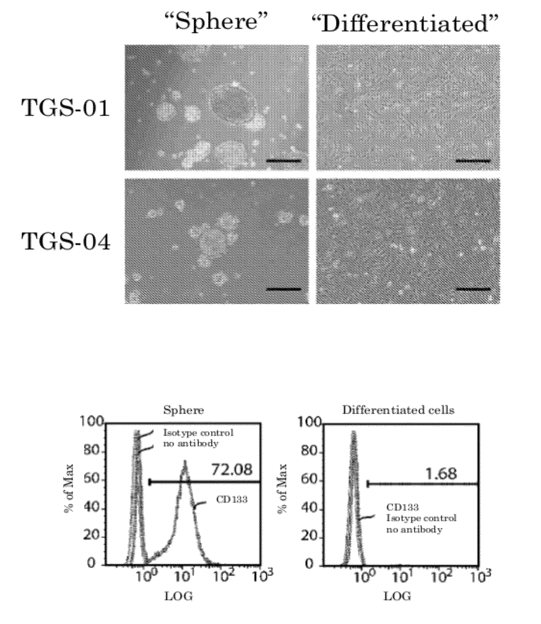
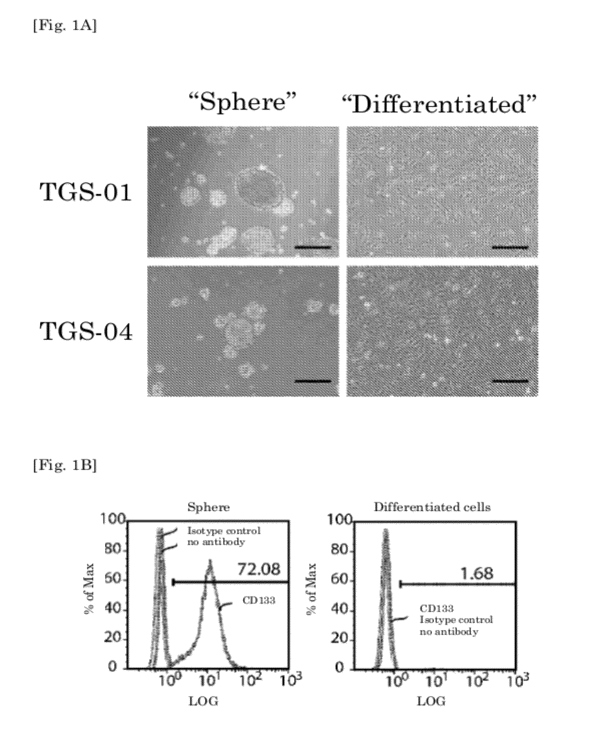
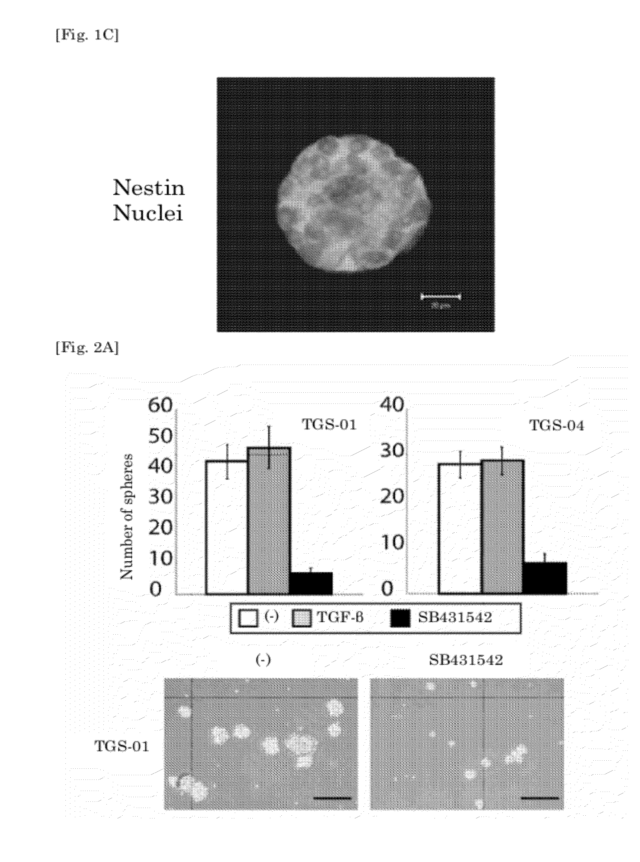
[0037]FIG. 1A is a photo of glioma spheres and differentiated cells. The scale bar is 100 μm. FIG. 1B is the results of measuring the CD133-positive cells in the glioma spheres and differentiated cells using flow cytometry. FIG. 1C is a photo showing the expression of the neural precursor cell marker Nestin in the glioma spheres. The scale bar is 20 μm.
[0038]FIG. 2A and FIG. 2B are the results of a sphere formation assay in the presence of the TGF-β inhibitor SB431542. The scale bar is 100 μm. FIG. 2C is the result of a sphere formation assay in the presence of a TGF-β receptor II / Fc chimera. The scale bar is 100 μm. FIG. 2D is the result of a sphere formation assay in the presence of the TGF-β inhibitor A-78-03 or LY364947. FIG. 2E is the result of a sphere formation assay in the presence of the TGF-β signal-negative regulatory factor SMAD7. FIG. 2F is a photo showing that the spheres, once formed, become adhesion cells when SB431542 is added.
[0039]FIG. 3A is the result of examining the impact of a TGF-β inhibitor on the percentage of CD133-positive cells in the glioma spheres. FIG. 3B is the result of examining the expression of neural precursor cell markers or nerve cell differentiation markers in the glioma spheres in the presence of a TGF-β inhibitor.
[0040]FIG. 4A is the result of measuring the impact of TGF-β or SB431542 on the expression of Sox2 using quantitative real-time RT-PCR. FIG. 4B is the result of examining the impact on SMAD2 and SMAD3 by siRNA, and on Sox2 expression induction by TGF-β. FIG. 4C is the result of measuring the expression of Sox2 at the protein level in the presence of TGF-β or SB431542. FIG. 4D is the result of measuring the impact of Sox2 knockdown by siRNA on the sphere formation abilities of glioma stem cells. FIG. 4E and FIG. 4G are the results of the impact of Sox2 knockdown by siRNA on glioma stem cell self-renewal ability measured using limiting dilution assay. FIG. 4F is the result of the impact of Sox2 knockdown by siRNA on the percentage of CD133-positive cells in the glioma spheres measured using flow cytometry. FIG. 4H is the result of examining the impact of Sox2 knockdown by siRNA on the percentage of Nestin-positive cells and GFAP-positive cells among all the cells.
[0041]FIG. 5A is the result of measuring the sphere formation ability of the glioma stem cells that expressed excessive Sox2 using sphere formation ability assay in the presence or absence of a TGF-β inhibitor. FIG. 5B is the result of measuring the percentage of Nestin-positive cells or GFAP-positive cells in the glioma spheres that expressed excessive Sox2.

Problems solved by technology

Nevertheless, many brain tumors invade the surrounding brain tissue and spinal tissue to create areas where the tumor cells and normal brain tissue are mixed, so surgical resection is troublesome.
Radiotherapy also damages the surrounding normal tissue easily.
Further, as the blood-brain barrier exists between the blood and the brain, even if a drip is applied, there are therapeutic agents that do not reach the brain, and chemotherapy might not be effective.
As described above, treating brain cancers is generally difficult, and effective treatments for highly-malignant glioblastomas in particular are not to be seen.
Cancer treatments to date have targeted the tumor bulk, and it is possible that insufficient effects have been obtained against the cancer stem cells.

Method used


Image

  • Brain Tumor Stem Cell Differentiation Promoter, and Therapeutic Agent for Brain Tumor
  • Brain Tumor Stem Cell Differentiation Promoter, and Therapeutic Agent for Brain Tumor
  • Brain Tumor Stem Cell Differentiation Promoter, and Therapeutic Agent for Brain Tumor

Examples


implementation example 1

Regulation of Glioma Sphere and Differentiated Glioma Cells, and Evaluation of their Characteristics

[0129]To clarify the mechanism by which glioma stem cellularity is maintained, TGS-01 and TGS-04 were first cultured in serum-free culture as described above to obtain spheres.

[0130](See FIG. 1A Left.) Both had self-renewal abilities, and showed similar action to that of the original cancer when transplanted into the brains of immunosuppressed mice. On the other hand, when the same samples were cultivated in 10% fetal bovine serum, the spheres did not form. (See FIG. 1A Right.) Cells cultivated in this way are sometimes called “differentiated glioma cells” or “differentiated cells” below.

[0131]CD133 (Prominin-1) has been reported as a glioma-initiating cell marker. (See non-patent document 1.) Here, the cells were separated into single cells, and the CD133 expression levels measured using flow cytometry. The results are shown in FIG. 1B. An increase in the percentage of CD133-positive...

implementation example 3

Maintenance of Stem Cellularity in Glioma-Initiating Cells by Sox2, and the Induction of Sox2 Expression due to TGF-β

[0139]To clarify the mechanism by which the stem cellularity of glioma-initiating cells is maintained by TGF-β signals, the effects of TGF-β and 31542 on the expression of stem cell markers were examined.

[0140]The results are shown in FIG. 4A. The expression of Sox2 mRNA, which is a HMG-box factor, was induced 24 hours after processing using TGF-β (100 μM), but when using SB431542 (1 μg), Sox2 expression was suppressed after 24 hours, and the low expression level continued subsequently for seven days.

[0141]In contrast, the expression of other pluripotent stem cell-related molecules such as Oct-4, NANOG, and LIF, etc., were not greatly affected by processing using TGF-β or an inhibitor. (See FIG. 14.) Further, it has been reported that NANOG and LIF are induced in many cells by stimulating TGF-β. (Xu et al., Cell Stem Cell, 2008, 3: 206; Bruna et al., Cancer Cell, 2007...

implementation example 4

Effects of TGF-β Inhibitors on Cells that Over-Express Sox2

[0148]To investigate more deeply the role of Sox2 in maintaining stem cell cellularity due to TGF-β, adenovirus coated with Sox2 cDNA was stained, and the effects of TGF-β inhibition on glioma-initiating cells that had been made to over-express Sox2 was examined.

[0149]Even if SB431542 was administered to glioma-initiating cells that had been made to over-express Sox2, sphere formation abilities were reduced only slightly compared to cells that had been made to over-express LacZ. (See FIG. 5A.) Further, even if SB431542 was administered to cells that over-expressed Sox2, from the fact that the percentage of Nestin-positive cells did not fall, and the percentage of GFAP-positive cells did not increase (see FIG. 5A), it was determined that glioma cells maintain stem cellularity.

[0150]From these results, it was understood that loss of stem cellularity using TGF-β inhibitors was due to the down-regulation of Sox2, which maintains...


PUM

PropertyMeasurementUnit
Therapeuticaaaaaaaaaa
Levelaaaaaaaaaa

Abstract

A promoter for differentiation of brain tumor initiating cells is provided.

Description

RELATED APPLICATION DATA[0001]This application is a continuation of PCT application number PCT / JP2010 / 051950 designating the United States and filed Feb. 10, 2010; which claims the benefit of U.S. Provisional application No. 61 / 151,645 and filed Feb. 11, 2009 both of which are hereby incorporated by reference in their entireties.TECHNICAL FIELDS[0002]This invention concerns a differentiation promoter for brain tumor stem cells, which reduces the tumorigenicity by promoting the differentiation of brain tumor stem cells.BACKGROUND TECHNOLOGY[0003]Brain tumor is the general name for tumors that occur in subcranial tissue, and which occur not only in the brain cells, but in all subcranial tissues, such as the dura, arachnoid membrane, subcranial blood vessels, and peripheral nerves, etc. Tumors in the brain require treatment that that does not damage as far as possible the normal surrounding tissue. Nevertheless, many brain tumors invade the surrounding brain tissue and spinal tissue to...

Claims


Application Information

Patent Timeline
IPC IPC(8): A61K49/00A61K31/7088A61K31/711A61K31/713A61P35/00C12Q1/02C12Q1/68C40B30/02C07H21/00C07H21/02G01N33/53
CPCA61K31/4439A61K31/713C12N2310/14C12N15/113C12N2310/11A61K45/06A61P35/00A61P43/00A61K48/00
Inventor MIYAZONO, KOHEIIKUSHIMA, HIROAKIMIYAZAWA, KEIJITODO, TOMOKIINO, YASUSHI
Owner THE UNIV OF TOKYO
Learn More