

Method and a Kit To Detect Malignant Tumors and Provide a Prognosis
a kit and malignant tumor technology, applied in the field of cancer diagnosis and prognosis, can solve the problems of low efficiency of currently available methods for obtaining specific exosome preparations, and achieve the effect of improving existing clinical tests
- Summary
- Abstract
- Description
- Claims
- Application Information
AI Technical Summary
Benefits of technology
Problems solved by technology
Method used
Image

Examples
example 1
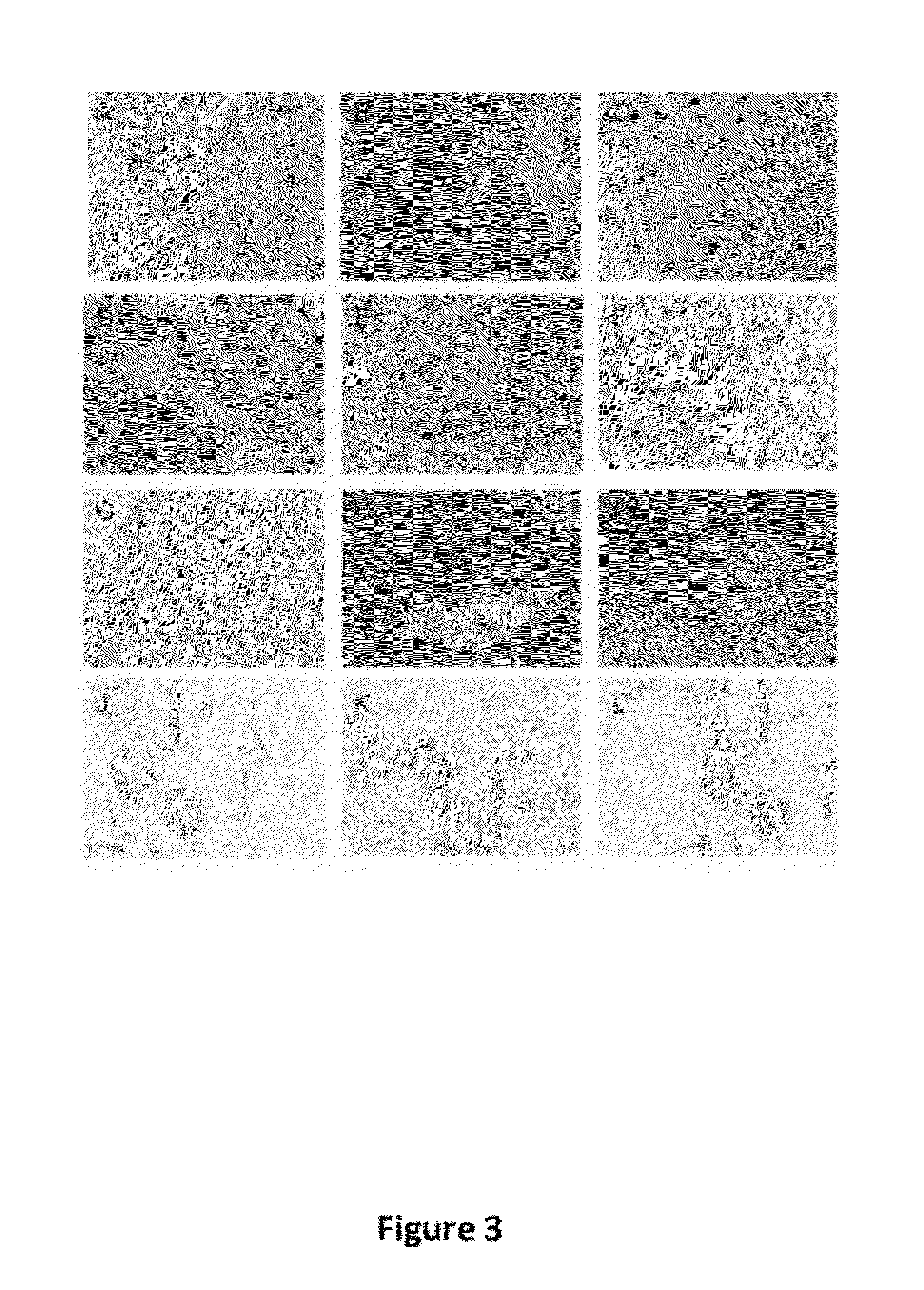
Immunocytochmemistry Shows TM9SF4 Protein Expression in Melanoma Cells
[0095]Immunocytochemistry and immunohistochemistry: For immunocytochemistry melanoma cells and macrophages, cultured on glass chamber slides (Falcon), and PBL, cytospun on glass slides, were fixed with 80% methanol 10 minutes at 4° C. and stained for TUCAP-1, TUCAP-1 mouse serum or preimmune control serum. Malignant melanoma and corresponding normal skin tissue from Biomax array slides (Biomax) were immunostained with pre-immune serum, for anti-TUCAP-1 mouse antiserum. Melanoma was also stained for anti-gp100 (Immunotech) while normal skin was also stained for anti-ezrin (Sigma). Proteins were visualized using the peroxidase antiperoxidase method in single staining (Dako) and counterstained with Mayer's hematoxylin.
[0096]FIG. 3A-C shows that MM2 cell lines (A), Peripheral blood lymphocytes (B), and in vitro differentiated Macrophages (C), were negative for mouse preimmune serum. However, malignant melanoma culture...
example 2
Western Blot, Immunofluorescence and FACSs Studies Show TM9SF4 (TUCAP-1)-Protein Expressing in Colon Carcinoma Cells
[0097]Purification of exosomes purification from cell culture supernatants and plasma. Supernatants from human cell lines were harvested from 72 hours 70-75% confluent cell cultures, and exosomes were isolated as follows. Briefly, after centrifugation of cells at 300 g for 10 minutes, supernatants were centrifuged at 1,200 g for 20 minutes followed by 10,000 g for 30 minutes. Supernatants were filtered using a 0.22 μm filter (Millipore Corp., Bedford, Mass.) and centrifuged at 100,000 g for 1 h in an ultracentrifuge (Sorval) in order to pellet exosomes. Exosomes were washed and resuspended in PBS.
[0098]Western Blotting
[0099]Cell lysates were prepared from cells harvested at 70-90% confluency. Cell flasks were washed with PBS, cells detached with Trypsin-EDTA for 2 minutes at room temperature and trypsin quenched with cell growth media containing 5% serum. Cells were wa...
example 3
RT-PCR Analysis Shows TUCAP-1 Protein to be Expressed in Prostate Cancer, Osterocarcoma, B Cell Lymphoma, Breast Carcinoma, and Ovary Carcinoma Cell Lines
[0102]PCR analysis. Expression of Tucap-1 transcripts was assessed by RT-PCR on several tumor cell lines. Total RNA from the cells was obtained by the RNAzoI (Invitrogen) method and RNA templates were used for RT-PCR amplification.
[0103]Primers for TUCAP-I detection were:
(SEQ ID NO: 9)tgtgtgaaacaagcgccttc,and(SEQ ID NO: 10)atgaggtggacgtagtagt.
[0104]These primers amplify a fragment of 349 base pairs.
[0105]Primers to detect GAPDH were:
(SEQ ID NO: 11)ccatggagaaggctggggand(SEQ ID NO: 12)caaagttgtcatggatgacc.
[0106]The suggested feature of TUCAP-1 as a tumor marker is strongly supported by a reported expression of a corresponding mRNA in a wider panel of human malignant cancer cell lines including B lymphoma, breast carcinoma, prostate cancer and ovary carcinoma, as is demonstraded on FIG. 6.
PUM
 Abstract
Description
Claims
Application Information