Yeast cell wall particles for receptor-targeted nanoparticle delivery

a technology of receptor-targeted nanoparticles and yeast cells, which is applied in the direction of genetic material ingredients, instruments, microcapsules, etc., can solve the problems of limited technology, manufacturing challenges, and small size of yeast cells, and achieve effective drug delivery systems

Active Publication Date: 2012-03-22
UNIV OF MASSACHUSETTS
View PDF0 Cites 41 Cited by
  • Summary
  • Abstract
  • Description
  • Claims
  • Application Information

AI Technical Summary

Problems solved by technology

While macroscopic drug delivery systems such as transdermal patches, implantable osmotic pumps and implantable subcutaneous depots (e.g., NORPLANT™) have had some success, these technologies are often limited in, for example, achieving controlled dosing, compatibility with a variety of chemically distinct biomolecules, discomfort associated with administration and the like.
While the high surface area / volume ratio of microcapsules, microspheres and liposomes favor the release of the active agent, their small size provides challenges in manufacturing.
The development of effective drug delivery systems presents multiple challenges, such as issues of drug solubility, targeting, in vivo stability and clearance, and toxicity.
A primary challenge to realizing the full promise of nanoparticle based drug delivery is the lack of optimal strategies to achieve selective and efficient cellular targeting.
505-515, 2007), and several competing uptake mechanisms result in undesired processes including off-target accumulation in other organs tissues and cells, rapid clearance from in vivo circulation (especially nanoparticles less than 5 nm) (S. V. Vinogradov, et al., Advanced Drug Delivery Reviews, vol.

Method used


Image

  • Yeast cell wall particles for receptor-targeted nanoparticle delivery
  • Yeast cell wall particles for receptor-targeted nanoparticle delivery
  • Yeast cell wall particles for receptor-targeted nanoparticle delivery

Examples


example 1

Test Ability of Biotinylated Qdots to Bind to Biotinylated YGP Surface and to GP Encapsulated Biotinylated Nanocores

[0181]Experiments were conducted to test the ability of loading quantum dots (CDSe / ZnS quantum dots) into GP by affinity trapping or on the surface of the shell using biotin and streptavidin affinity interactions.

[0182]To prepare YGP tRNA / PEI-biotin cores, 1 mg of YGP was mixed with 6 μL 10 mg / mL yeast RNA (Sigma), and incubated 2 hours at room temperature. Then 250 μL of 0.1% PEI biotin was added and the particles allowed to swell for 30 min, then sonicated and incubated 1 hour at room temperature. The particles were collected by centrifugation, washed three times with 500 μL 0.9% saline and resuspended in 500 μL PBS. The particles were sonicated, particle number counted using a hemacytometer, a 1×10+8 / mL suspension prepared in PBS and stored frozen at −20C.

[0183]To bind streptavidin Q-Dots to YGP-biotin shells and YGP tRNA / PEI-biotin cores PBS, particles and Qdots we...

example 2

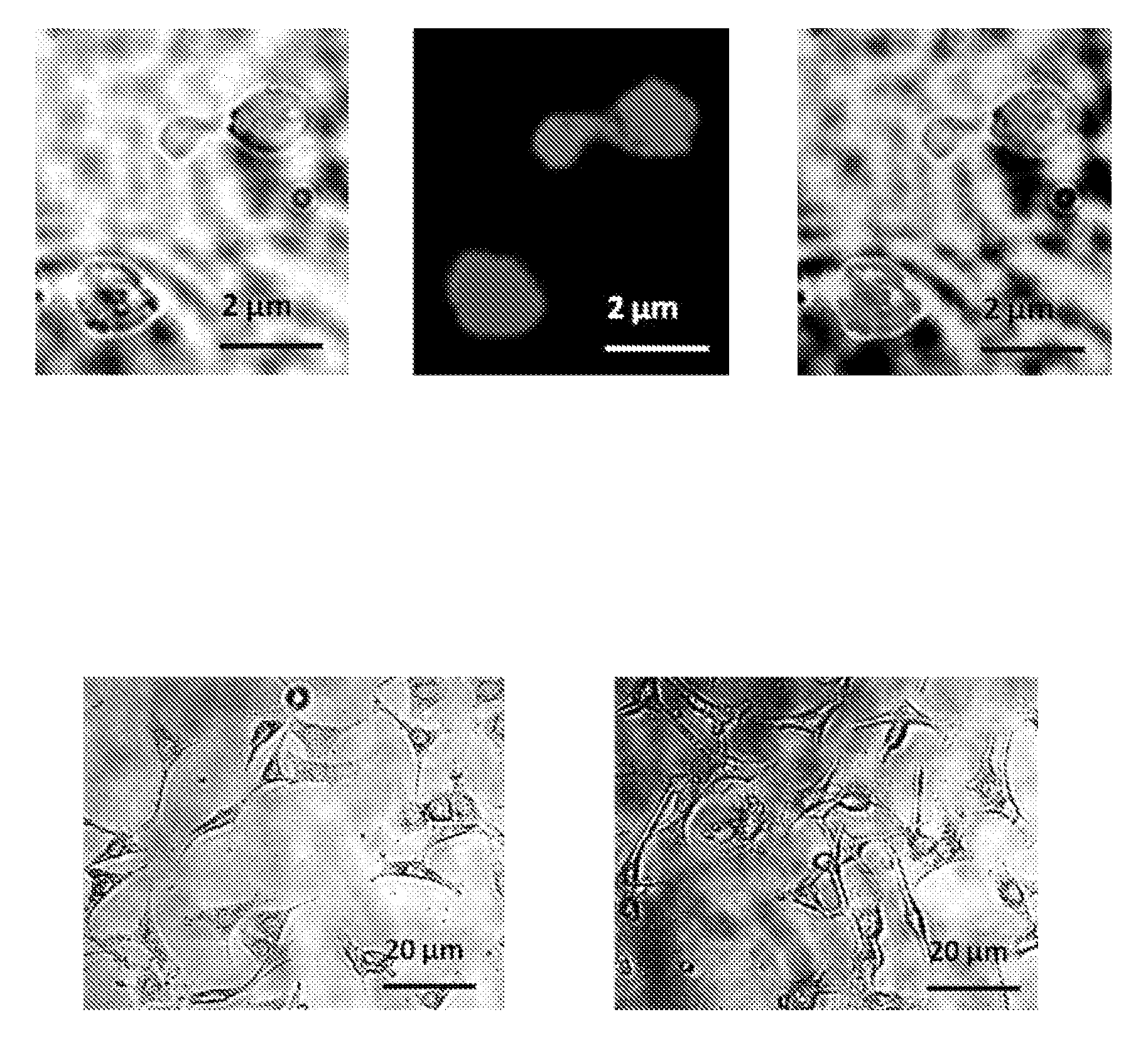
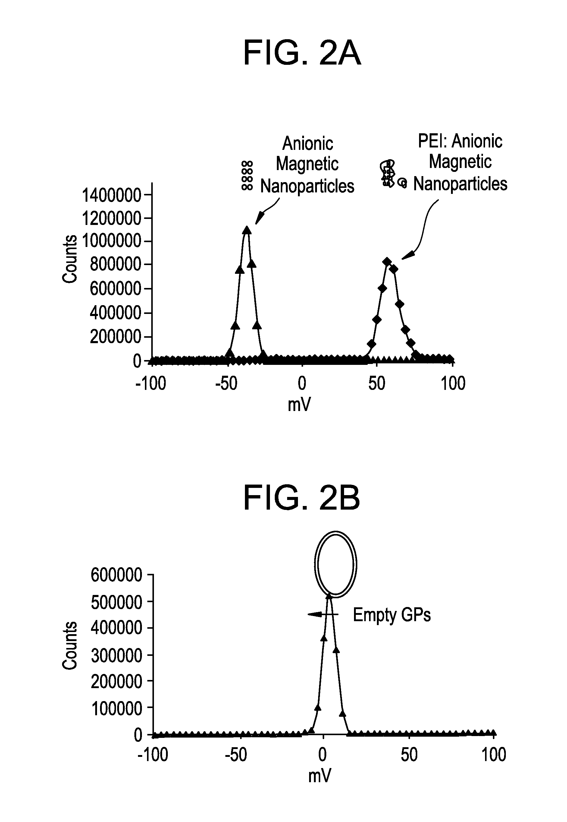
Loading of GP with Magnetic Nanoparticles

[0186]Experiments were conducted to show that magnetic NPs could be loaded into or onto GPs. To make GP formulations loaded with magnetic nanoparticles tubes containing 1 mg or 100 mg of empty GPs were loaded with 5 μL of magnetic Nanoparticles / mg GP. Cationic 10 nm magnetic nanoparticles (EMG 607) or anionic 10 nm magnetic nanoparticles (EMG 707) were loaded (Ferrotec, Bedford, N.H.). Following incubation for 2 hr at room temperature tubes were frozen and lyophilized. A water push reaction was carried out with 5 μl water / mg GPs at room temperature for >1 hours. The tubes were then frozen and lyophilized, and contents resuspended in 70% ethanol for 15 minutes. Free nanoparticles were separated from GP magnetic nanoparticles by centrifugation and supernatants were collected and optical density measured to determine unencapsulated magnetic nanoparticles. The 70% ethanol washing steps were repeated twice and the pellets lyophilized. The pellets ...

example 3

GP Trapping of Au / MUA Nanoparticles

[0189]Gold nanoparticles coated with NanoXact™ Gold nanoparticles from Nanocomposix (San Diego, Calif.) (Table 3; 250 uL, 0.02 mg Au / mL) were incubated with 11-mercaptoundecanoic acid (MUA, 110 uL of a 0.6 mg / mL solution). The final volume was adjusted to 500 uL with 0.01 M NaOH. The samples were then incubated overnight at room temperature. The MUA derivatized gold nanoparticles (Au / MUA) were centrifuged for 20 min at 10000 rpm. The supernatant was carefully removed and the pellets were washed twice with 1 mM NaCl. The washed pellets were resuspended in 250 uL of 1 mM NaCl and OD measurement at 525 nm was used to calculate % of nanoparticle recovery.

TABLE 3TubeAu NP size1Au 10 nm2Au 30 nm3Au 40 nm4Au 50 nm5Au 100 nm

[0190]GP Loading of Au / MUA Nanoparticles—

[0191]Empty glucan particles were mixed with Au / MUA nanoparticles (5 uL of nanoparticle suspension / mg GP) as indicated in Table 4. The pellet was mixed and incubated at room temperature for 1 hou...


PUM

PropertyMeasurementUnit
diameteraaaaaaaaaa
diameteraaaaaaaaaa
diameteraaaaaaaaaa

Abstract

The present invention generally relates to yeast cell wall microparticles loaded with nanoparticles for receptor-targeted nanoparticle delivery. In particular, the present invention relates to trapping nanoparticles either on the surface or inside a yeast glucan particles, for example, yeast glucal particles. The present invention further relates to methods of making the yeast cell wall particles loaded with nanoparticles. The present invention also relates to methods of using the yeast cell wall particles loaded with nanoparticles for receptor-targeted delivery of the nanoparticles, e.g., drug containing nanoparticles.

Description

RELATED APPLICATIONS[0001]This application claims priority to U.S. Provisional Patent Application No. 61 / 373,828, filed Aug. 14, 2010. The entire content of the foregoing application is incorporated herein by reference.BACKGROUND OF THE INVENTION[0002]Drug delivery systems are designed to provide a biocompatible reservoir of an active agent for the controlled release of the active agent dependent either on time, or on local conditions, such as pH. While macroscopic drug delivery systems such as transdermal patches, implantable osmotic pumps and implantable subcutaneous depots (e.g., NORPLANT™) have had some success, these technologies are often limited in, for example, achieving controlled dosing, compatibility with a variety of chemically distinct biomolecules, discomfort associated with administration and the like. Accordingly, there has been continuing interest in developing microscopic drug delivery systems aimed at overcoming some of the limitations inherent in the above descri...

Claims


Application Information

Patent Timeline
Patent Type & Authority Applications(United States)
IPC IPC(8): A61K9/14A61K49/00B82Y5/00
CPCA61K9/5068A61K9/5115B82Y5/00A61K47/489A61K47/4893A61K47/48853A61K47/52A61K47/61A61K47/6921A61K47/6933A61K47/6941A61K31/704A61K35/66A61K9/1652A61K9/1682A61K48/00G01N33/587
Inventor OSTROFF, GARY R.SOTO, ERNESTO
Owner UNIV OF MASSACHUSETTS
Why Eureka
Try Eureka