Radiographic image detector and gain setting method for radiographic image detector

a radiographic image and detector technology, applied in the direction of radio control devices, instruments, television systems, etc., can solve the problems of affecting the image correction process, the density gradation of the subsequent image is not adequately reproduced,

Active Publication Date: 2012-08-09
FUJIFILM CORP
View PDF0 Cites 34 Cited by
  • Summary
  • Abstract
  • Description
  • Claims
  • Application Information

AI Technical Summary

Benefits of technology

[0012]I view of the foregoing, an object of the present invention is to provide a solution for acquiring high-quality radiographic images with properly reproduced density gradations without complicating the processing for image correction after the A / D conversion.
[0029]According to the present invention, after a radiation dose, before reading the signal charges, a dose profile representative of at least linear distribution of the dosed amounts of radioactive rays is measured to determine the gain based on the contrast in the dose profile. Then the signal charges are read out from the pixels at the determined gain. Thus, high-quality radiographic images may be acquired with properly reproduced density gradations without complicating the image processing after the A / D conversion.

Problems solved by technology

As a result, it sometimes happens that the subsequent image does not adequately reproduce the density gradation.
Therefore, the second method will complicate the image correcting processes after the A / D conversion.

Method used


Image

  • Radiographic image detector and gain setting method for radiographic image detector
  • Radiographic image detector and gain setting method for radiographic image detector
  • Radiographic image detector and gain setting method for radiographic image detector

Examples


Embodiment Construction

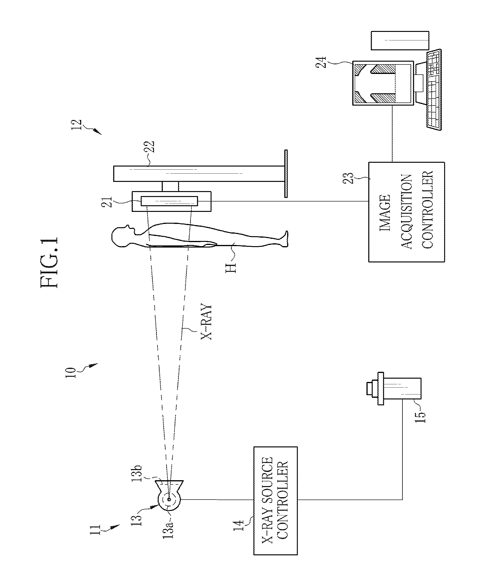
[0043]In FIG. 1, an x-ray radiography system 10 consists of an x-ray projector 11 and radiographic equipment 12. The x-ray projector 11 consists of an x-ray source 13, an x-ray source controller 14 for controlling the x-ray source 13, and an activator switch 15. The x-ray source 13 has an x-ray tube 13a for radiating x-rays and a collimator 13b for limiting the radiation field of x-rays from the x-ray tube 13a.

[0044]The x-ray tube 13a has a cathode which includes a filament for emitting thermions and an anode (target) against which the thermions strike to radiate x-rays. The collimator 13b may for example be made of lead plates shielding x-rays, which are put together in a double-cross formation to form a center aperture for letting x-rays pass through it. The lead plates are movable to change the size of the center aperture so as to limit the radiation field to a suitable range.

[0045]The x-ray source controller 14 includes a high voltage generator for supplying a high voltage to t...


PUM


Abstract

In a radiography system, after a radiation dose, signal charges are accumulated in sensor pixels in an imaging area of a flat panel detector corresponding to the dosed amounts of radioactive rays on the pixels. The signal charges are read out from the pixels and converted into voltage signals representative of density levels of respective pixels of a radiographic image. Before reading the signal charges, a dose profile representative of distribution of the dosed amounts of radioactive rays is measured by leak currents from the pixels or bias currents through bias lines for applying a bias voltage to the pixels. Based on the contrast or difference between the maximum value and the minimum value in the dose profile, a gain of amplifiers for the voltage signals is determined such that the gain increases with decreasing contrast.

Description

BACKGROUND OF THE INVENTION[0001]1. Field of the Invention[0002]The present invention relates to a radiographic image detector and a gain setting method for the radiographic image detector.[0003]2. Description of the Related Art[0004]In the medical field, radiography systems utilizing radioactive rays such as x-rays for imaging are known. An x-ray radiography system includes an x-ray projector having an x-ray source for radiating x-rays, and radiographic equipment that receives x-rays after having been projected from the x-ray projector toward a subject and penetrating through the subject, thereby to acquire a radiograph or x-ray image that represents information on the subject. As the radiographic equipment, an x-ray image detector using a flat panel detector (FPD) in place of conventional x-ray film or an imaging plate (IP) has recently been developed and used in practice, which can output digital data of an acquired x-ray image, as disclosed in JPA 2009-219538.[0005]As described ...

Claims


Application Information

Patent Timeline
Patent Type & Authority Applications(United States)
IPC IPC(8): G01T1/24
CPCH04N5/32H04N5/3653H04N5/361H04N25/672H04N25/63
Inventor KONDOU, KIYOSHI
Owner FUJIFILM CORP
Why Eureka
Try Eureka