Klf family members regulate intrinsic axon regeneration ability

a technology of intrinsic axon and klf family, which is applied in the direction of antibody medical ingredients, peptide/protein ingredients, metabolic disorders, etc., can solve the problems of regeneration failure, developmental loss of intrinsic axon growth capacity of neurons,

Inactive Publication Date: 2012-09-06
UNIV OF MIAMI
View PDF1 Cites 6 Cited by
  • Summary
  • Abstract
  • Description
  • Claims
  • Application Information

AI Technical Summary

Benefits of technology

[0082]The dose of an agent of the invention, or composition thereof, administered to an animal, particularly a human, in the context of the present invention should be sufficient to effect at least a detectable amount of a therapeutic response in the individual over a reasonable time frame (e.g., a CNS axon-regeneration effective amount). The exact amount of the dose will vary from subject to subject, depending on the species, age, weight and general condition of the subject, the severity or mechanism of any disorder being treated, the particular agent or vehicle used, its mode of administration and the like. The dose used to achieve a desired effect in vivo will be determined by the potency of the particular agent employed, the pharmacodynamics associated with the agent in the host, the severity of the disease state of infected individuals, as well as, in the case of systemic administration, the body weight and age of the individual. The size of the dose also will be determined by the existence of any adverse side effects that may accompany the particular inhibitory agent, or composition thereof, employed. It is generally desirable, whenever possible, to keep adverse side effects to a minimum.
[0083]Dosages for administration of an inhibitory or stimulatory agent of the invention can be in unit dosage form, such as a tablet or capsule. The term “unit dosage form” as used herein refers to physically discrete units suitable as unitary dosages for human and animal subjects, each unit containing a predetermined quantity of an inhibitor of the invention, alone or in combination with other suitable therapeutic agents, calculated in an amount sufficient to produce the desired effect in association with a pharmaceutically acceptable diluent, carrier, or vehicle.
[0084]The specifications for the unit dosage forms of the present invention depend on the particular agent of the invention, or composition thereof, employed and the effect to be achieved, as well as the pharmacodynamics associated with each polypeptide, or composition thereof, in the host. In some embodiments, the dose administered is a “CNS axon-regeneration effective amount.”
[0085]One skilled in the art can easily determine the appropriate dose, schedule, and method of administration for the exact formulation of the composition being used, in order to achieve the desired response in the individual patient.

Problems solved by technology

In addition to the development of an inhibitory CNS environment, a developmental loss in neurons' intrinsic capacity for axon growth is thought to contribute to regeneration failure.
However, manipulating these regulators of axon growth, even when simultaneously overcoming environmental inhibition, only partially restores regeneration, suggesting that additional intrinsic axon growth regulators remain to be identified.

Method used


Image

  • Klf family members regulate intrinsic axon regeneration ability
  • Klf family members regulate intrinsic axon regeneration ability
  • Klf family members regulate intrinsic axon regeneration ability

Examples


example i

Materials and Methods

Constructs for Transfection

[0098]For the screen in hippocampal neurons, constructs in pEXPRESS-1, pSPORT or pCMV-SPORT6 (Open Biosystems) were co-transfected with pMAX (EGFP, Amaxa), and compared to an empty vector / pMAX co-transfection. The purchased constructs for the screen were full-length rat cDNAs (19 / 111, 17%), or else mouse (73%) or human (10%) when the full-length rat cDNA was unavailable.

[0099]Flag-tagged KLF4 constructs in a CS2+ vector were a generous gift of Chunming Liu (Univ of Texas). The flag control vector was purchased from Genecopoeia. Mouse KLF4 (Open Biosystems) and mCherry (gift of Roger Tsien, UCSD) were cloned into the pIRES2-eGFP vector (Clontech).

[0100]KLFs -1, -4, -5, -6, -7, -10, -12, -15, and -17 were obtained from Open Biosystems. KLF2 was a kind gift from Jerry Lingrel, Univ. of Cincinnati. The open reading frame of KLF9 was cloned from postnatal rat cortex, and KLFs -3, -8, -11, -13, -14, and -16 were cloned from mouse spleen or t...

example ii

KLF Family Members Regulate Intrinsic Axon Regeneration Ability

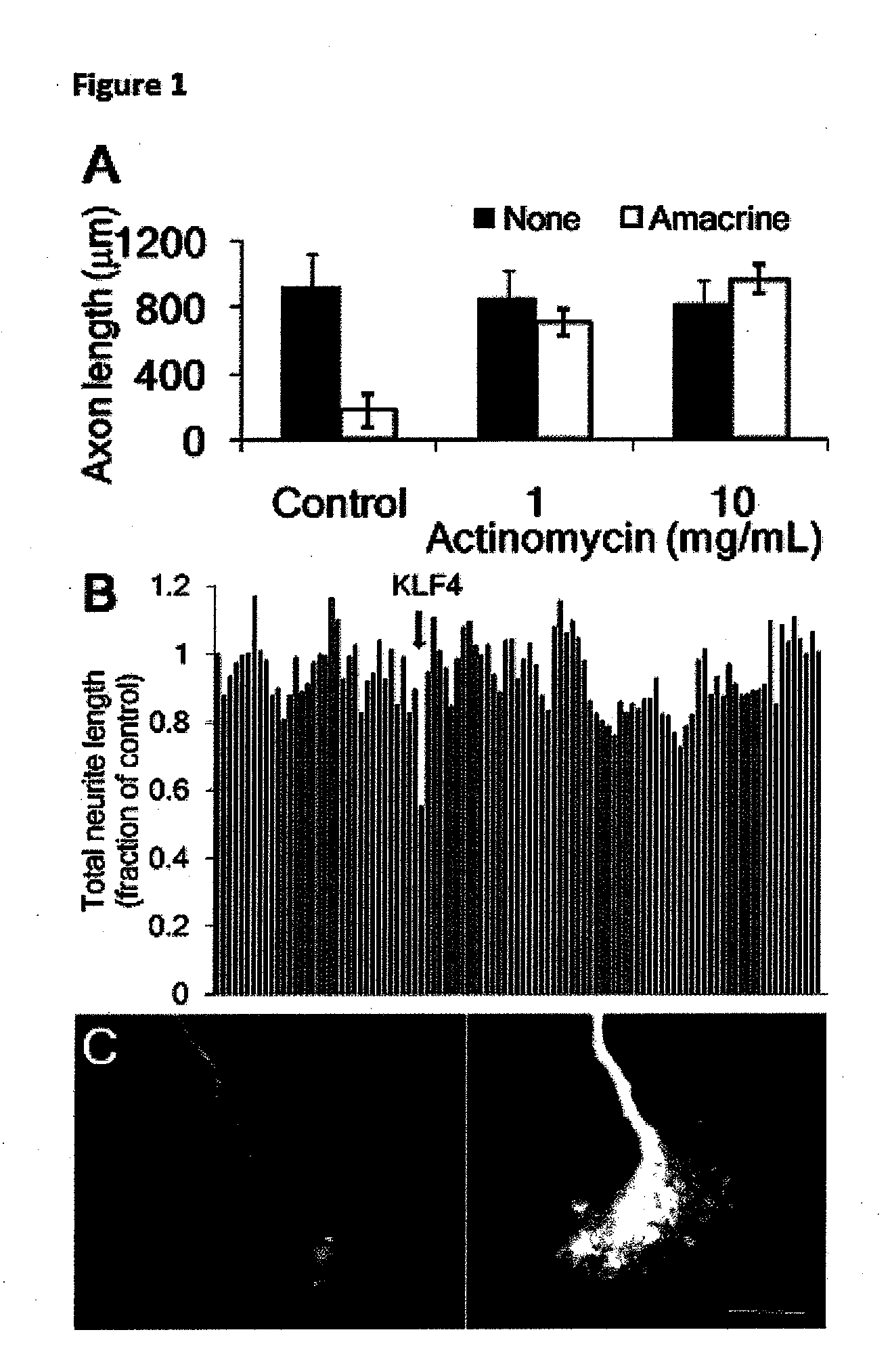
[0127]To investigate the molecular basis for the developmental loss of axon growth ability in RGCs, we took advantage of the fact that co-culture with amacrine cell membranes is sufficient to signal embryonic RGCs to decrease their rapid axon growth (Goldberg et al. (2002), supra). Addition of the transcriptional inhibitor actinomycin D blocked this effect of amacrine membranes, and embryonic RGCs retained their capacity for axon growth (FIG. 1A). These data suggest that gene transcription is required for the developmental loss of intrinsic axon growth ability in RGCs.

[0128]To identify candidate genes, we profiled gene expression from embryonic day 17 (E17) through postnatal day 21 (P21) RGCs (Wang et al. (2007), supra), spanning the period when axon growth ability declines in vivo (Goldberg et al. (2002), supra; Chen et al. (1997), supra). We screened 111 candidates whose expression changed greater than 3-fold by overex...

example iii

Further Experiments Showing that KLF Family Members Regulate Intrinsic Axon Regeneration Ability

1. KLF4 Overexpression in Hippocampal Neurons Decreases Neurite Growth and Neurite Initiation.

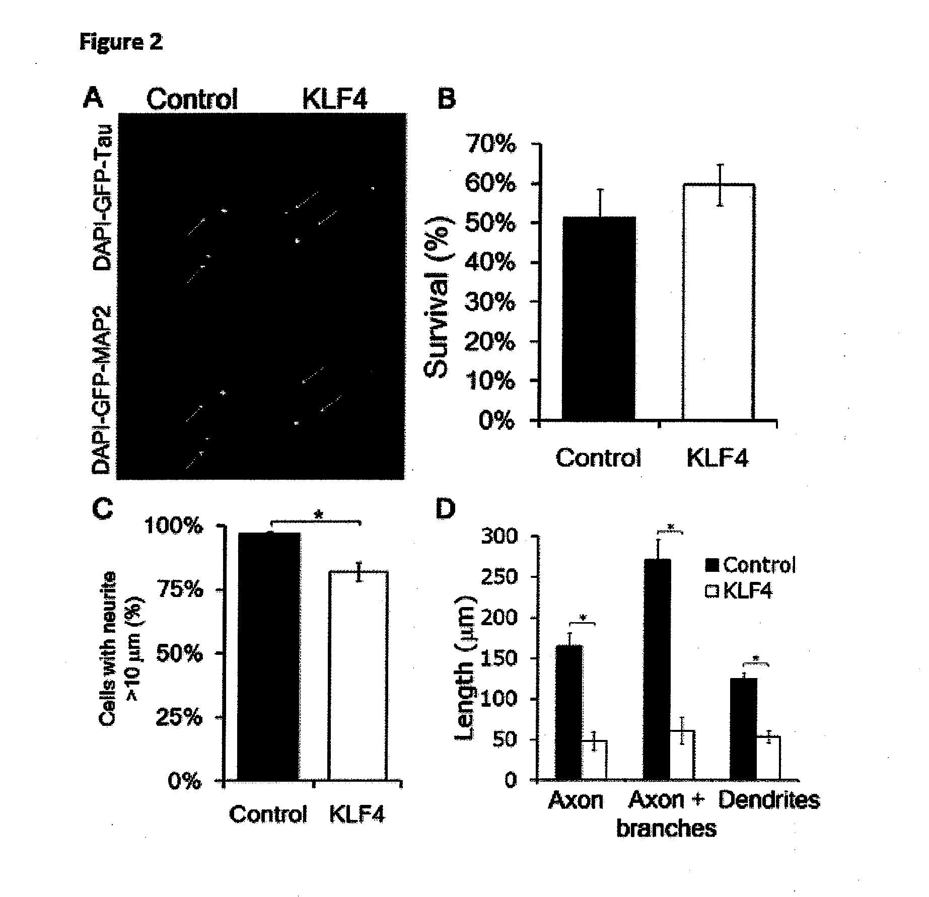
[0136]The results of this study are shown in FIG. 7. A-D) E18 hippocampal neurons were co-transfected with KLF4 or control plus EGFP, cultured on laminin-coated plates, and immunostained for Tau (neurites) and MAP2 (dendrites). A) There was no difference in survival by nuclear morphology and DAPI intensity between control- and KLF4-transfected neurons (Mean±SD). B) Transfected EGFP+ cells (arrows) were imaged to detect DAPI, EGFP, and either Tau (top) or MAP2 (bottom). KLF4-transfected neurons had shorter axons and dendrites. (Scale bar, 50 μm) C) KLF4 overexpression decreased the percentage of transfected neurons that were able to extend at least 1 neurite >10 μm (N=5; *p<0.01, paired t-test; mean±SEM). D) KLF4 overexpression decreased both axon (Tau+ / MAP2−) and dendrite (MAP2+) length (*p<0.01,...


PUM

PropertyMeasurementUnit
pHaaaaaaaaaa
temperatureaaaaaaaaaa
length analysisaaaaaaaaaa

Abstract

This invention relates, e.g., to a method for promoting CNS axon regeneration, comprising (1) inhibiting the expression or activity in a neuron of one or more of the members of the Krüppel-like transcription factor (KLF) family that suppress axon growth (e.g., KLF 1, 2, 3, 4, 5, 9, 12, 13, 14, 15 and/or 16), and/or (2) stimulating the expression or activity in a neuron of one or more of the members of the KLF family that promote axon growth (e.g., KLF 6 and/or 7).

Description

[0001]This application claims the benefit of the filing date of U.S. Provisional Application 61 / 239,873, filed Sep. 4, 2009, which is incorporated by reference in its entirety herein.[0002]The research leading to this invention was supported in part by an R01grant, number EY 016790, from the National Eye Institute (NEI); an R03 grant, number NS061348, from the National Institute of Neurological Disorders and Stroke (NINDS); a P30 grant, number EY014801, from the NEI; and NINDS training grants T32 NS07492 and T32 NS007459. The Government of the United States of America has certain rights in the invention.BACKGROUND INFORMATION[0003]Adult mammalian central nervous system (CNS) axons are unable to regenerate after injury, but immature CNS neurons regenerate axons robustly. In addition to the development of an inhibitory CNS environment, a developmental loss in neurons' intrinsic capacity for axon growth is thought to contribute to regeneration failure. For example, after birth, axonal ...

Claims


Application Information

Patent Timeline
Patent Type & Authority Applications(United States)
IPC IPC(8): A61K48/00A61P25/00A61K39/395C12Q1/68C12N5/0793A61K38/02
CPCC07K14/4702C12N2501/60C12N5/0619A61P25/00
Inventor GOLDBERG, JEFFREY L.LEMMON, VANCE P.BIXBY, JOHNMOORE, DARCIEBLACKMORE, MURRAY
Owner UNIV OF MIAMI
Why Eureka
Try Eureka