Method and Apparatus for Cervical Cancer Screening

a cervical cancer and autofluorescence technology, applied in the field of cervical cancer screening systems and methods, can solve the problems of limited pathology resources in under-developed low sensitivity of pap smear, and inability to meet the needs of patients in resource-poor regions of the world, so as to reduce the overall cost or size of the system, and reduce the overall cost and cost of the overall apparatus.

Inactive Publication Date: 2012-09-13
WELLER BROPHY LAURA ANN
View PDF16 Cites 11 Cited by
  • Summary
  • Abstract
  • Description
  • Claims
  • Application Information

AI Technical Summary

Benefits of technology

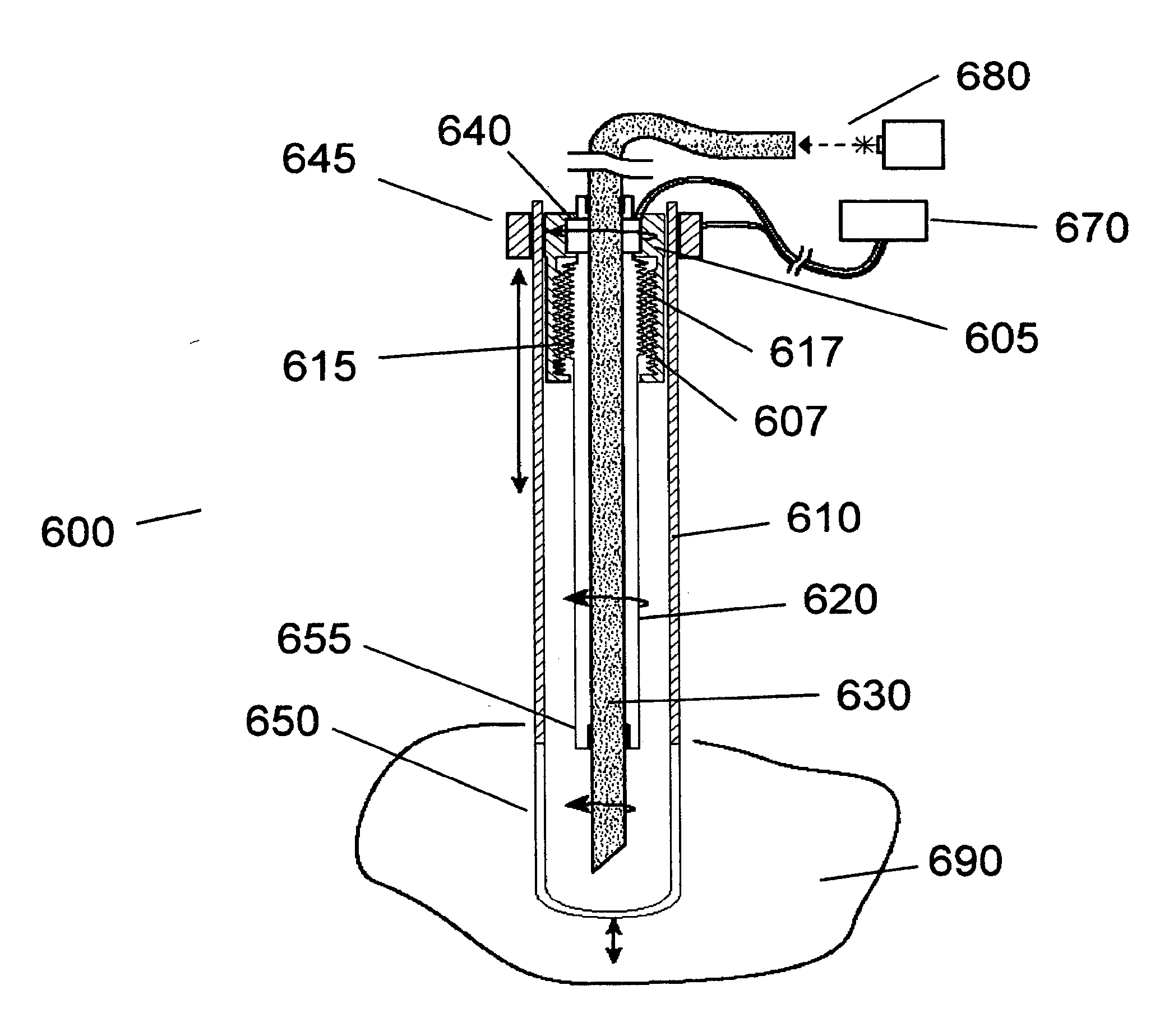
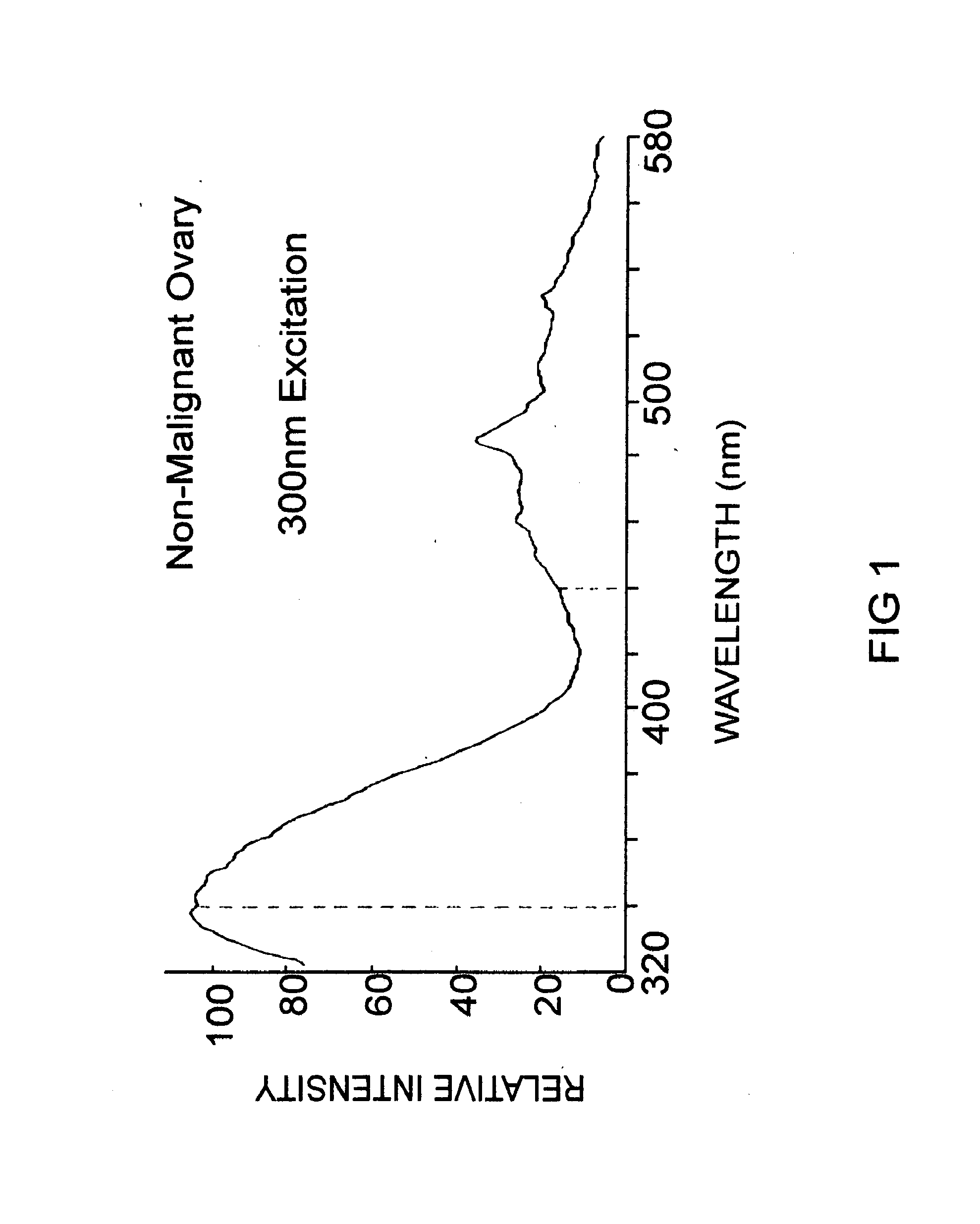
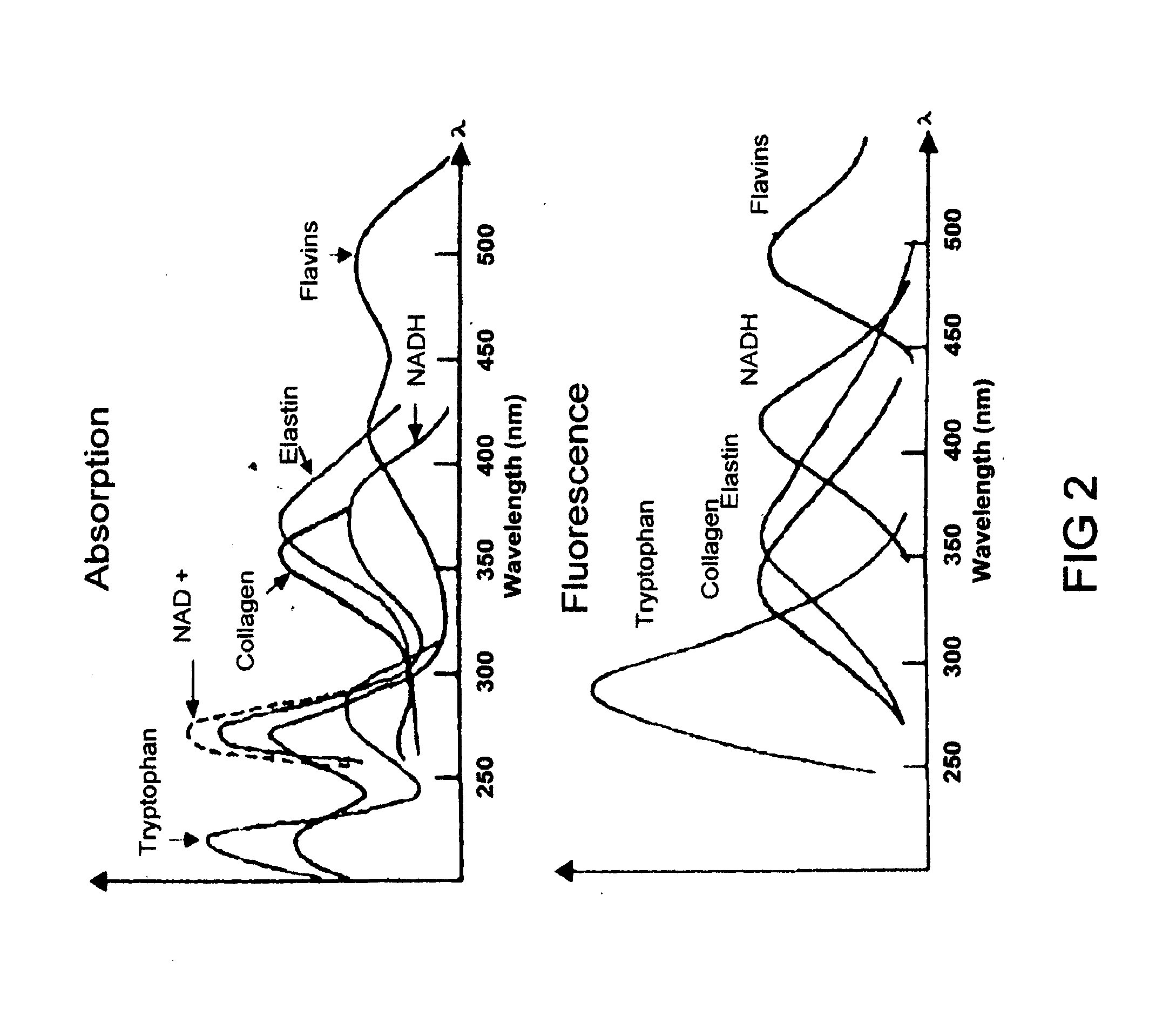
[0044]The present invention includes several advantages, not all of which are incorporated in a single embodiment. The present cervical cancer screening method and apparatus offers both sensitivity and specificity >80%, and provides immediate results so that treatment may be offered at the time of the test. The present cervical cancer screening method and apparatus fully screens both the surface of the cervix and the endocervical canal, wherein both the surface of the cervix and the endocervical canal are interrogated through illumination, for example, in the approximately 290-310 nm band. This band is selected in some embodiments, as it has been shown to produce fluorescence correlated with cervical cancer disease state and to reduce confounding fluorescence signals from inflamed, but otherwise normal cervical tissues. In addition, the components of the imaging and cervical canal probe systems are integrated or shared to reduce overall cost or size of the system. Unlike conventional Pap smears, HPV DNA tests, and colposcopic evaluations, the present cervical cancer screening method and apparatus offers an immediate result, enabling the patient to receive follow-on care at the same visit, if necessary. The present cervical cancer screening method and apparatus eliminate the need for separate pathological confirmation of disease, although such confirmation may be used in conjunction with the present method, if desired. The present invention is advantaged over other fluorescence-based methods in that it preferentially uses the 290-310 nm illumination band, interrogates and screens both the surface of the cervix and the cervical canal, incorporates a means to control the position of the probe in the cervical canal, can control the point-wise and total exposure of the cervical canal to illumination light, and includes an apparatus that provides for the sharing of components so to reduce overall apparatus size and cost.

Problems solved by technology

While use of Pap smears has produced a 74% drop in cervical cancer deaths in the US, there are limitations.
Pap smear sensitivity is low, ˜51%, and false negative rates for Pap tests range from 34-63% mainly due to inadequacies of sampling, slide preparation, or processing problems.
An added limitation of current methods of cervical cancer screening using Pap smears is the limited availability of pathology resources in under-developed regions of the world.
While cervical cytologic testing has significantly reduced the cervical cancer death rate in developed countries, it remains unavailable in resource-poor regions of the world.
In resource-poor regions of the world, the death rate results from the lack of cytologic screening facilities, insufficient numbers of trained personnel, and inability of patients to travel repeatedly to health facilities for the screening follow-up.
Colposcopy and fluorescence imaging cannot be modified readily to allow simultaneous imaging of the surface of the cervical canal and of the surface of the cervix.
A large, fixed optical viewing system, such as a colposcope, designed to image the surface of the cervix, and not configured to be inserted into the cervical canal, will not provide the required interrogation of the cervical canal.
In cases where the transformation zone has regressed either partially or fully into the cervical canal, current colposcopy and fluorescence methods will not be successful in the complete screening of the cervix for cancers and precancers.
No commercial methods are available today that include imaging and light-based interrogation of the cervical canal.
In addition, the probes proposed for use in the endocervical canal have not controlled the per-point and total exposure of the illumination light.
The inventors do not disclose a means to use rotation and translation with a fiber-based device, nor do they address how the probe motion may be automated so to control the exposure of the tissue under test.
The authors do not teach the use of separately optimized probes for assessing a tubular structure and separately imaging an outer surface of that structure.
In the case of the analysis of cervical cancer, and the need to assess both the relatively narrow cervical canal as well as the outer surface of the cervix, the small tubular probe required to fit into the cervical canal would not be adequately large to enclose the imaging optics required to take a high quality image of the cervical surface.
Accordingly, these currently methods do not provide immediate results, require multiple collection and preparation steps for each sample, and depend upon trained pathology specialists to provide review of the pathology status.
Given the multiple collection and preparation steps, as well as the human interpretation of the samples, errors may occur, with sensitivity of the Pap smear method about 50%.
A low specificity means that the method may suffer from a significant number of false positive results, indicating that a patient has cervical cancer or precursors of cervical cancer when, in fact, their tissues are normal.
False positive results lead to over-treatment of patients, unnecessary medical procedures, and health risks.
In resource-poor countries, where trained pathologists and local health care facilities are rare, women do not have access to Pap smears.
Current fluorescence methods, however, are limited by relatively low specificity due to illumination wavelengths that excite confounding tissue fluorescence from inflamed tissue sites.
In addition, current methods and equipment fail to interrogate and screen both the outer surface of the cervix as well as the cervical canal.

Method used


Image

  • Method and Apparatus for Cervical Cancer Screening
  • Method and Apparatus for Cervical Cancer Screening
  • Method and Apparatus for Cervical Cancer Screening

Examples


examples

[0104]Table 1 compares existing technologies that detect the threshold of high-grade cervical neoplasias (CIN 2, CIN 3, CIS, and Invasive Cancer). Included in the table are experimental methods based upon cervical tissue fluorescence. The Guided Therapeutics data are from FDA trials. The 300 nm data are from published reports, and represent the results anticipated for a BioProbe according to the current invention.

TABLE 1Summary of methods for the screening and diagnosis ofcervical cancer, together with the Guided TherapeuticsLightTouch ™ currently in FDA trials, and the300 nm Fluorescence Excitation method proposed herein.ScreeningImmediateMethodSensitivitySpecificityResult?Turn-AroundPap Smear151%97%No1-2 weeksColposcopy396%48%No2-4 weeksfor biopsiesPap + HPV195%27%No1-2 weeksGuided95%55%YesNoneTherapeutics2Pap + Guided95%65%No1-2 weeksTherapeutics1300 nm88-97%   88-97%   YesNoneFluorescenceExcitation (Invitro)4-51Werner, c., W Griffith III, R Ashfaq, D Gossett, et. al., “Compariso...


PUM


Abstract

The present invention relates to an apparatus for cervical cancer screening, comprising one or more light sources aligned with a beginning of a first optical test path and a beginning of a second optical test path, one or more optical detectors aligned with an end of the first optical test path and an end of the second optical test path, and a processor coupled to the one or more light sources and the one or more optical detectors and methods for using the same. The present invention further relates to a method for cervical cancer screening.

Description

FIELD OF THE INVENTION[0001]The claimed invention relates generally to cervical cancer screening systems and methods, specifically to a light-based system and method for cervical cancer screening, and more specifically to an autofluorescence-based system and method for cervical cancer screening. Even more specifically, the invention pertains to an autofluorescence-based system and method for cervical cancer screening that illuminates tissues in certain embodiments at approximately the 290-310 nm band and includes screening of the cervical canal.BACKGROUND OF THE INVENTION[0002]In the 60 years since its development, the Pap test has become the most common means for the screening of cervical neoplasia. The method's success is due to the qualities of low cost, minimal invasiveness, and ease of collection of the cervical cell sample. While use of Pap smears has produced a 74% drop in cervical cancer deaths in the US, there are limitations.[0003]Pap smear sensitivity is low, ˜51%, and fa...

Claims


Application Information

Patent Timeline
Patent Type & Authority Applications(United States)
IPC IPC(8): A61B6/00
CPCA61B1/00165A61B1/043A61B1/303A61B5/0062A61B2562/0233A61B5/0084A61B5/4331A61B5/6852A61B5/0071Y02A90/10
Inventor WELLER-BROPHY, LAURA ANN
Owner WELLER BROPHY LAURA ANN
Why Eureka
Try Eureka