
Methods and means to alter lipid biosynthesis by targeting multiple enzymes to suborganelle domains
a technology of suborganelle domains and enzymes, applied in biochemistry apparatus and processes, peptides, enzymology, etc., can solve the problems of defective prior art, insufficient seed yield of these plants, and produced fatty acids or lipids
- Summary
- Abstract
- Description
- Claims
- Application Information
AI Technical Summary
Problems solved by technology
Method used
Image

Examples
example 1
Materials and Methods
[0112]For all methods sterile pipette tips and reaction tubes were used. All solutions were set up with sterile water (ddH2O).
[0113]Hardware / Equipment
[0114]ABI PRISM® 3100 Genetic Analyzer Applied Biosystems, Foster, USA
[0115]Automatic TLC Sampler 4 Camag, Berlin, Germany
[0116]Centrifuge 5810 R Eppendorf AG, Hamburg, Germany
[0117]Centrifuge 5415 D Eppendorf AG, Hamburg, Germany
[0118]Centrifuge 5417 R Eppendorf AG, Hamburg, Germany
[0119]Chromatogramm Immersion Devise III Camag, Berlin, Germany
[0120]ColorView II 3.3 MegaPixel CCD Camera Olympus, Hamburg, Germany
[0121]Gel-Detection System DIANA Raytest, Straubenhardt, Germany
[0122]Gel-Detection System AIDA Raytest, Straubenhardt, Germany
[0123]Glass beads Roth, Karlsruhe, Germany
[0124]Gold particles BioRad, Hercules, USA
[0125]Mastercycler personal Eppendorf, Wesseling-Berzdorf, Germany
[0126]Olympus BX51 Olympus, Hamburg, Germany
[0127]Olympus U-RFL-T Olympus, Hamburg, Germany
[0128]PDS1000 / He Biolistic Particle Delive...
example 2
Plant and Yeast Expression Constructs Used Herein.
[0231]To perform retargeting experiments, truncated versions of MmFAR1 were generated to determine which domains are essential for peroxisomal localization and which further domains can retarget the MmFAR enzymes from the peroxisomes to the cytosol or ER.
[0232]mCherry is a red fluorescent protein and its excitation and emission maxima are 587 nm and 610 nm, respectively. Expression of fusion proteins with mCherry enables to determine their localization in vivo by fluorescence microscopy. mCherry was cloned by PCR amplification into the BamHI and EcoRI sites (5′ and 3′ of mCherry, respectively) of the pUC18-ENTRY or the pESC-URA plasmids. The full length and the truncated open reading frames (ORFs) of MmFAR1 were modified using the Phusion Polymerase (Finnzymes, Espoo, Finland) and a standard PCR protocol with the respective primers listed in Materials and methods, introducing an EcoRI restriction site at the 5′ prime end and a XhoI r...
example 3
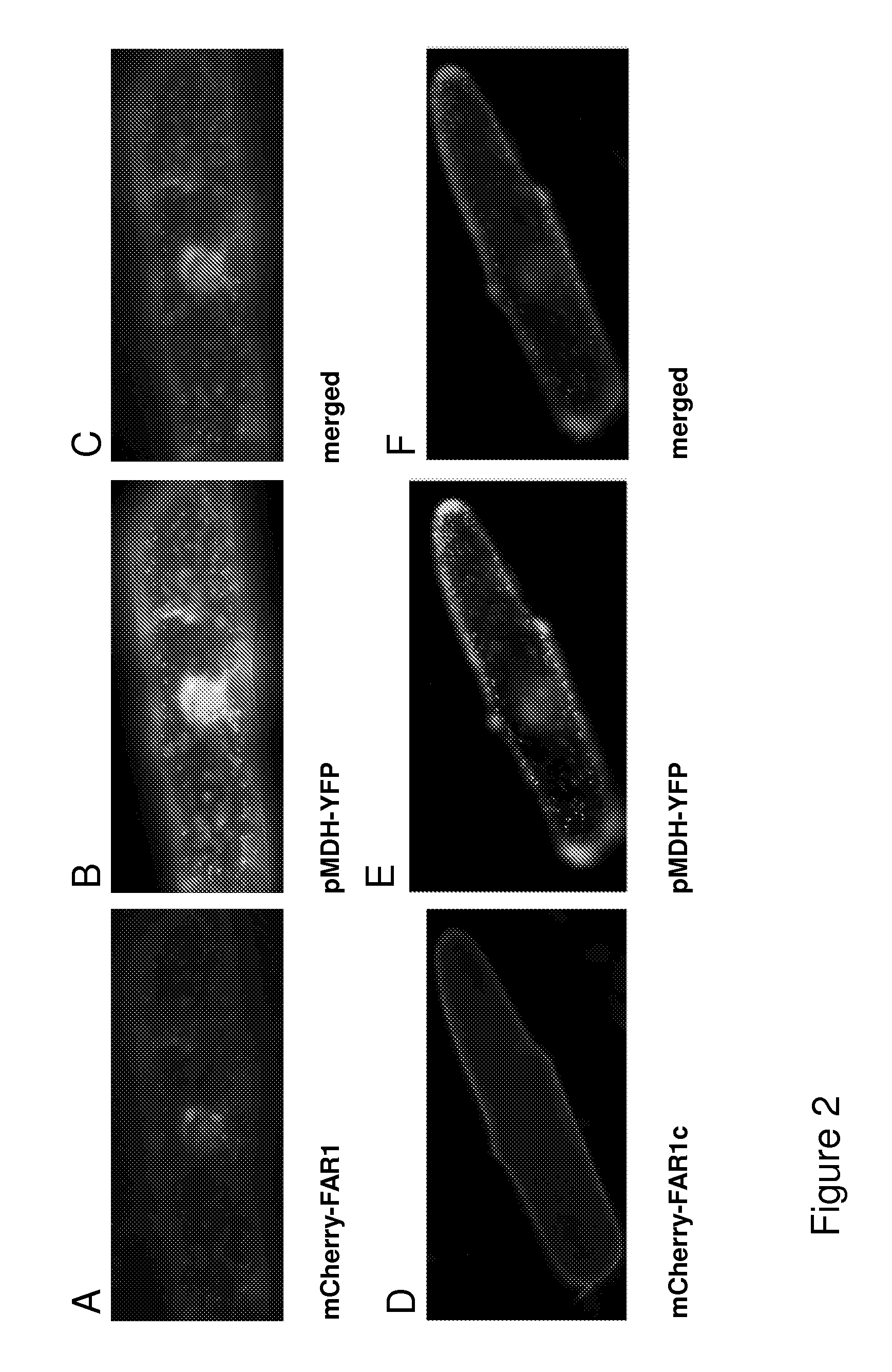
Subcellular Localization of MmFar1 and truncated MmFar1c and Fusion Proteins to Oleosin by Transient Expression in Onion Epidermis Cells.
[0235]The fusion- or the truncated mCherry-MmFAR1 constructs, arranged in the pUC18-ENTRY vector, were transferred into the plant expression vector pCAMBIA3300 by clonase reaction. After selection, the positive clones were precipitated on gold particles along with a peroxisomal marker (MDH). Those particles were then shot in onion cells in order of transient transformation. After incubation over night the transformed onion cells were examined via fluorescent microscopy.
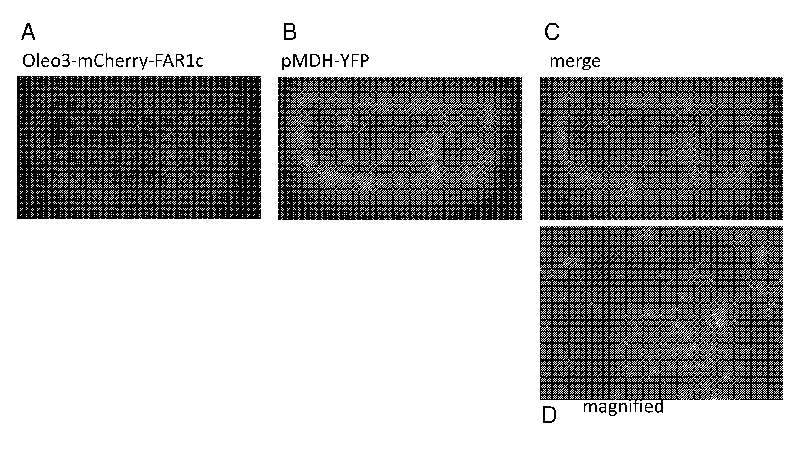
[0236]FIG. 2 shows the localization of the diverse MmFAR1 constructs tagged to mCherry. Images A of FIG. 2 shows onion epidermis cells expressing the mCherry-MmFAR1 construct. The fluorescence of mCherry-MmFAR1 was detected in the cytoplasm as small puncta that co-localized with the peroxisomal marker protein MDH (FIG. 2 B). observation corresponds to the described peroxisomal locali...
PUM
| Property | Measurement | Unit |
|---|---|---|
| Fraction | aaaaa | aaaaa |
| Acidity | aaaaa | aaaaa |
Abstract
Description
Claims
Application Information