
Apparatus and Method of Non-Contrast Magnetic Resonance Angiography of Abdominal and Pelvic Arteries

a technology of magnetic resonance angiography and abdominal and pelvis arteries, which is applied in the field of medical imaging, can solve the problems of difficult nc mra of the abdominopelvic arteries and is likely not well-suited for the abdomen, and achieve the effect of substantially focusing on the tissu

Inactive Publication Date: 2012-11-22
NEW YORK UNIV
View PDF3 Cites 22 Cited by
  • Summary
  • Abstract
  • Description
  • Claims
  • Application Information

AI Technical Summary

Benefits of technology

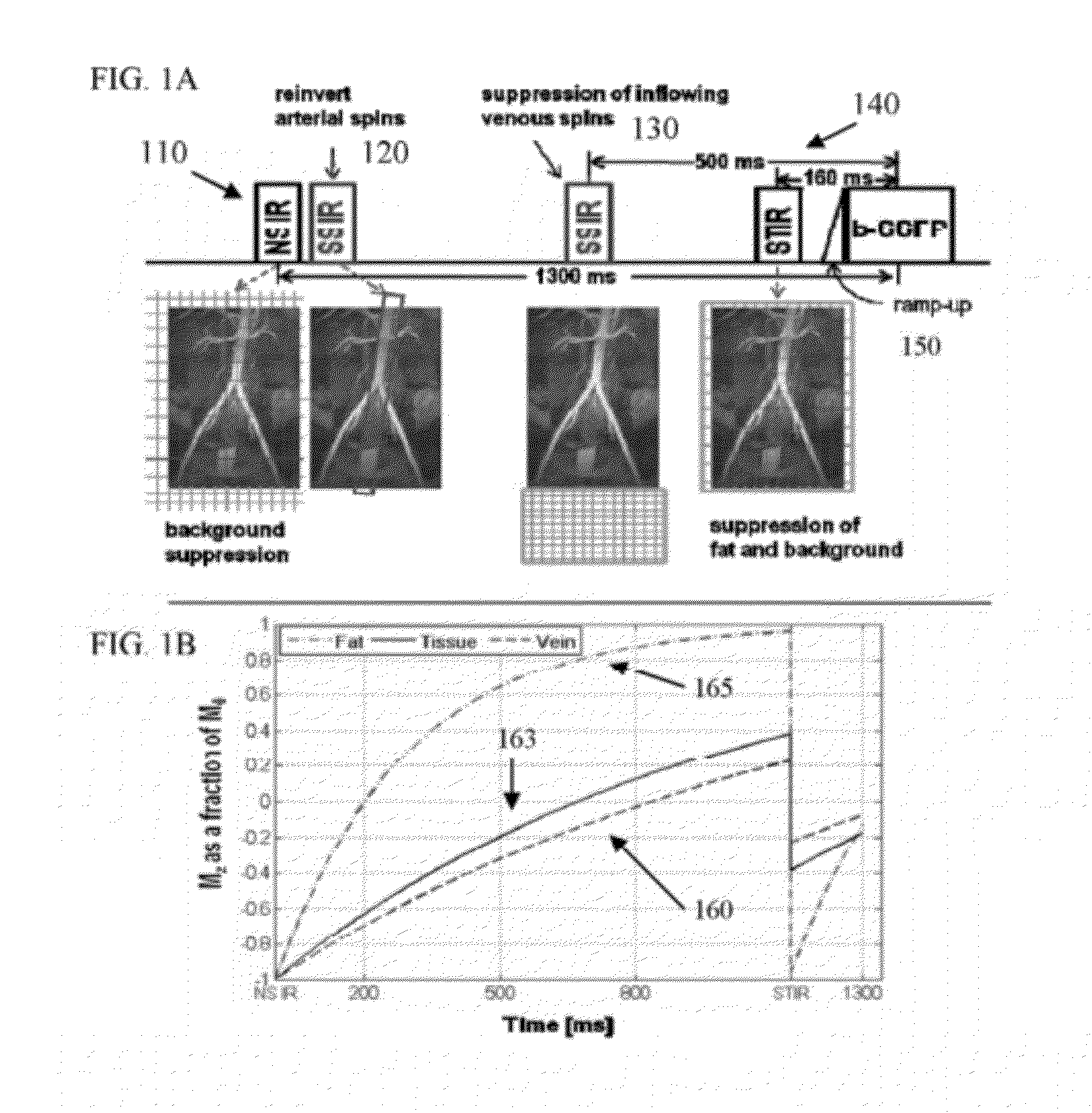
[0011]The tissue(s) can include an aortic bifurcation, and the element can include blood traveling through the aortic bifurcation. The RF pulses can include at least one first nonselective IR RF pulse configured to invert a longitudinal magnetization of at least one background tissue. The identifier can be established by re-inverting a longitudinal magnetization of the element associated with the tissue(s). The RF pulses can include a graphically oriented slice-selective RF pulse configured to re-invert the longitudinal magnetization of magnetic spins of the element associated with the tissue(s). A graphical orientation of the graphically oriented slice-selective RF pulse can be received, and the information can be generated using the graphical orientation. The graphical orientation can facilitate substantially focusing on the tissue(s) and exclude background tissues. The RF pulses can include a graphically oriented further slice-selective RF pulse configured to invert a longitudinal magnetization of an additional tissue. A graphical orientation of the graphically oriented further slice-selective RF pulse can be received, and the information can be generated using the graphical orientation of the further slice-selective RF pulse.
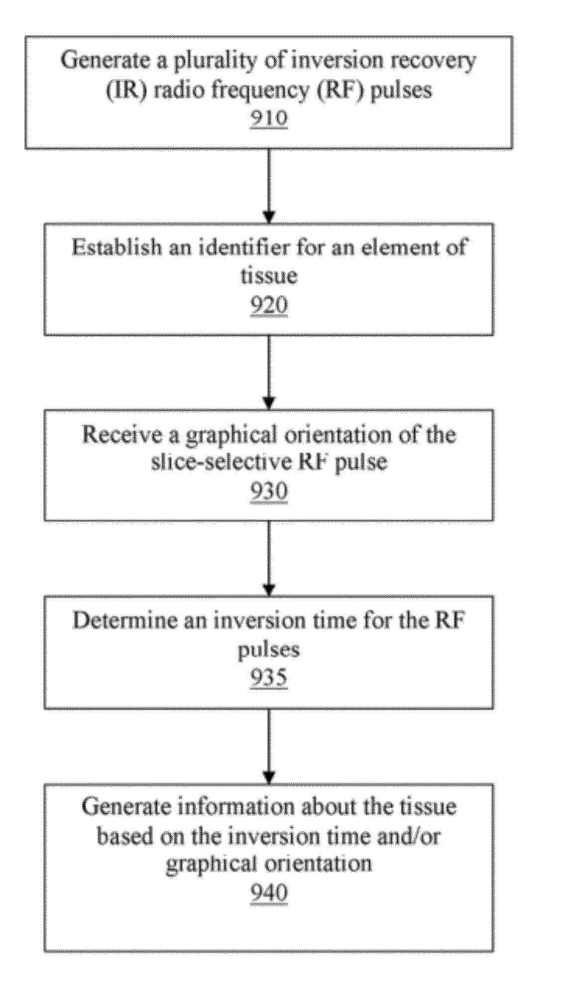
[0012]According to another exemplary embodiment of the present disclosure, the RF pulses can include a short inversion time inversion recovery (STIR) RF pulse configured to invert a longitudinal magnetization of the tissue(s)s within an imaging volume. The RF pulses can also include a graphically oriented slice-selective RF pulse configured to re-invert the longitudinal magnetization of magnetic spins of the element associated with the tissue(s) immediately following a first nonselective RF pulse which is configured to invert a longitudinal magnetization of background tissues. The tissue(s) can include an aortic bifurcation and the RF pulses can be IR pulses.

Problems solved by technology

However, recent safety concerns associated with gadolinium-based contrast agents (See Grobner T, Prischl F C. Gadolinium and nephrogenic systemic fibrosis.
NC MRA of the abdominopelvic arteries can be challenging due to a variety of factors including, e.g., respiratory motion suppression, large field of view (FOV) coverage, and depiction of vessels with variable hemodynamics and orientation.
J Magn Reson Imaging 2008; 28(1):181-189), but are likely not well-suited for the abdomen due to motion.
However, a single SS-IR b-SSFP MRA acquisition that can provide spatial coverage from renal arteries to iliac arteries with adequate background suppression has not been reported.

Method used


Image

  • Apparatus and Method of Non-Contrast Magnetic Resonance Angiography of Abdominal and Pelvic Arteries
  • Apparatus and Method of Non-Contrast Magnetic Resonance Angiography of Abdominal and Pelvic Arteries
  • Apparatus and Method of Non-Contrast Magnetic Resonance Angiography of Abdominal and Pelvic Arteries

Examples


Embodiment Construction

[0006]According to certain exemplary embodiments of the present disclosure, methods and apparatus can be provided which can facilitate / utilize a non-contrast MRA pulse sequence using 4 inversion-recovery (IR) pulses to provide coverage (e.g., a significant or complete coverage) from renal arteries to iliac arteries with a preferential background suppression. For example, the inversion times (TIs) and positions of the slice-selective IR pulses can be based on the Bloch equation governing T1 relaxation, T1 values of blood, tissue, and fat, and typical arterial blood flow rate. After pre-conditioning the magnetization with 4 IR pulses, a 3D b-SSFP readout can be used to image with bright arterial contrast.

[0007]For example, in particular exemplary embodiments of the present disclosure, methods and apparatus can be provided which can facilitate / utilize a preferential arterial conspicuity from the renal to the distal external iliac arteries with optimal background suppression. For exampl...


PUM


Abstract

Exemplary method and apparatus can be provided for generating information regarding at least one tissue. Using such exemplary method and apparatus, it is possible to generate a plurality of inversion recovery (IR) radio frequency (RF) pulses that are configured to establish an identifier of an element associated with at least one portion of the tissue(s). It is also possible to determine an inversion time associated with at least one of the RF pulses. Further, it is possible to generate, e.g., with a computing arrangement, the information regarding the tissue(s) based on the inversion time.

Description

CROSS-REFERENCE TO RELATED APPLICATION(S)[0001]The present application relates to and claims priority from U.S. Provisional Patent Application No. 61 / 454,832 filed Mar. 21, 2011, the entire disclosure of which is incorporated herewith by reference.STATEMENT REGARDING FEDERALLY SPONSORED RESEARCH[0002]The subject matter of the present disclosure was developed, at least in part, using Government support under Grant No. HL092439, awarded by the National Institute of Health and Grant No. 0730143N, awarded by the American Heart Association. Accordingly, the Federal Government has certain rights in the subject disclosure.FIELD OF THE DISCLOSURE[0003]Exemplary embodiments of the present disclosure relate to medical imaging, and more specifically to method, system, and apparatus for providing non-contrast magnetic resonance angiography (MRA).BACKGROUND INFORMATION[0004]Non-invasive assessment of the aortoiliac and the renal arteries can be used for the management of renal artery stenosis, a...

Claims


Application Information

Patent Timeline
IPC IPC(8): A61B5/055
CPCA61B5/0263A61B5/055G01R33/5635G01R33/5607G01R33/5602
Inventor KIM, DANIELLEE, VIVANATANASOVA, ILIYANA
Owner NEW YORK UNIV
Learn More