Hypothesis Validation of Far Wall Brightness in Arterial Ultrasound

a far wall brightness and ultrasound technology, applied in the field of data processing and imaging system validation, can solve the problems of difficult automated imt methods, and difficult to achieve the effect of accurate and fast detection

Inactive Publication Date: 2012-12-13
ATHEROPOINT
View PDF2 Cites 7 Cited by
  • Summary
  • Abstract
  • Description
  • Claims
  • Application Information

AI Technical Summary

Problems solved by technology

These challenges have complicated IMT measurement using conventional systems.
Though conventional methods are generally suitable, conventional methods have certain problems related to accuracy, speed, and reliability.
Further, the automated IMT methods suffer from the challenge that they hypothesize that the far wall has the brightest intensity.
IMT estimation having a value close to 1 mm is a very challenging task in ultrasound images due to large numbers of variabilities, such as: poor contrast, orientation of the vessels, varying thickness, sudden fading of the contrast due to change in tissue density, presence of various plaque components in the intima wall such as fibrous muscles, lipids, calcium, hemorrhage, etc.
Under normal resolutions, a 1 mm thick med...

Method used


Image

  • Hypothesis Validation of Far Wall Brightness in Arterial Ultrasound
  • Hypothesis Validation of Far Wall Brightness in Arterial Ultrasound
  • Hypothesis Validation of Far Wall Brightness in Arterial Ultrasound

Examples


Embodiment Construction

[0029]In the following description, for purposes of explanation, numerous specific details are set forth in order to provide a thorough understanding of the various embodiments. It will be evident, however, to one of ordinary skill in the art that the various embodiments may be practiced without these specific details.

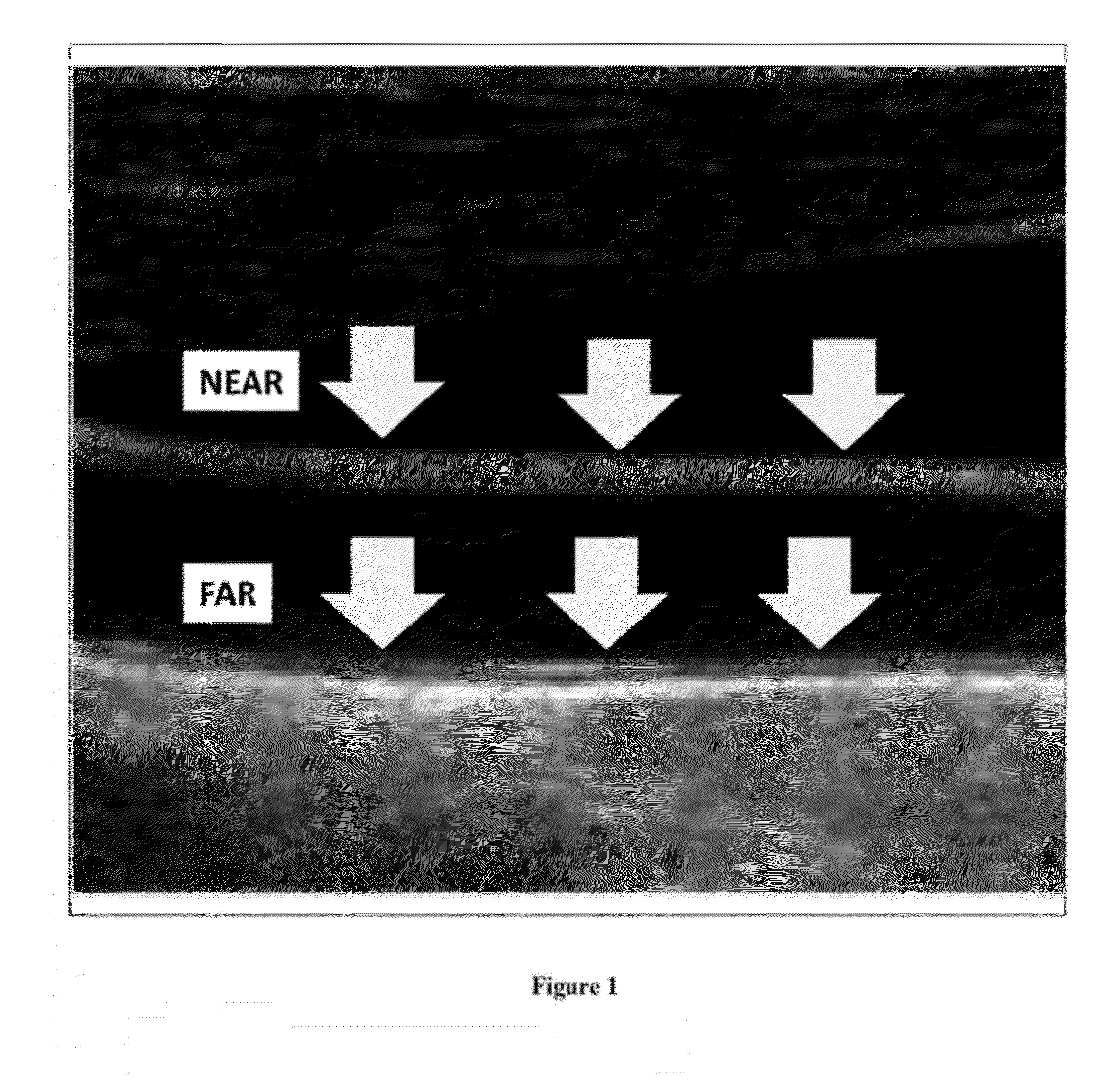
[0030]As explained above, an algorithm localized the adventitial wall based on the intensity local maxima of every column in the image, i.e., the far wall brightness compared to the near wall. Several systems developed for IMT measurements that were based on this hypothesis produced good agreement with expert segmentation. The assumption was based on manual intensity measurements on several, representative US images.

[0031]Thus, the assumption that the far wall brightness is the highest intensity in the image can be used as a basis for automatically finding the far adventitia borders and then automatically using that as a marker for IMT measurement. This application is ...


PUM


Abstract

Automated IMT system hypothesize that far wall of the common carotid artery has the highest intensity. In this current application, we verify that this hypothesis holds true for B-mode or RF-mode longitudinal ultrasound images of the carotid wall. The methodology consists of generating the composite image (arithmetic sum of images) from the database by first registering the carotid image frames with respect to a nearly straight carotid artery frame from the same database using (a) B-spline based non-rigid registration and (b) affine registration. Prior to registration, we segment the carotid artery lumen using a level set based algorithm followed by morphological image processing. The binary lumen images are registered and the transformations are applied to the original grayscale CCA images. These B-mode or RF-mode ultrasound images are then used for IMT computation using automated methods which hypothesize that far wall has the brightest intensity distribution.

Description

PRIORITY PATENT APPLICATIONS[0001]This is a continuation-in-part patent application of co-pending patent application Ser. No. 12 / 798,424; filed Apr. 2, 2010 by the same applicant. This is also a continuation-in-part patent application of co-pending patent application Ser. No. 12 / 799,177; filed Apr. 20, 2010 by the same applicant. This is also a continuation-in-part patent application of co-pending patent application. Ser. No. 12 / 799,558; filed Apr. 26, 2010 by the same applicant. This is also a continuation-in-part patent application of co-pending patent application Ser. No. 12 / 802,431; filed Jun. 7, 2010 by the same applicant. This is also a continuation-in-part patent application of co-pending patent application Ser. No. 12 / 896,875; filed Oct. 2, 2010 by the same applicant. This is also a continuation-in-part patent application of co-pending patent application Ser. No. 12 / 960,491; filed Dec. 4, 2010 by the same applicant. This is also a continuation-in-part patent application of c...

Claims


Application Information

Patent Timeline
IPC IPC(8): A61B8/13
CPCG06T7/0081G06T7/0091G06T7/602G06T2207/30101G06T2207/20036G06T2207/20161G06T2207/10132G06T7/11G06T7/155G06T7/62
Inventor SURI, JASJIT S.
Owner ATHEROPOINT
Why Eureka
Try Eureka