Systems, methods, compositions and devices for in vivo magnetic resonance imaging of lungs using perfluorinated gas mixtures

a technology of perfluorinated gas mixture and magnetic resonance imaging, which is applied in the field of noninvasive in vivo 19f magnetic resonance imaging, can solve the problems of low spirometry cost, lack of regional information about ventilation distribution or ventilation dynamics in the lung, and little appreciation of how to identify copd early

Active Publication Date: 2013-01-03
DUKE UNIV
View PDF3 Cites 15 Cited by
  • Summary
  • Abstract
  • Description
  • Claims
  • Application Information

AI Technical Summary

Benefits of technology

[0011]Embodiments of the invention can provide dynamic “free-breathing” cine images of a breathing lung (corresponding to a respiratory cycle). Some embodiments can provide dynamic images or pseudo-static images using gated image data collection over a plurality of respiratory cycles of a subject and reconstructed in a variety of manners to accommodate the respiratory cycle. Other embodiments employ short and / or long breath-hold techniques. Other embodiments include prospective or retrospective signal averaging of multiple image sets to improve the image quality (signal to noise and contrast to noise).
[0030]Optionally, the delivering step can be carried out using free-breathing, thereby allowing the subject to inhale and exhale the gas mixture over a plurality of respiratory cycles during the acquiring step.
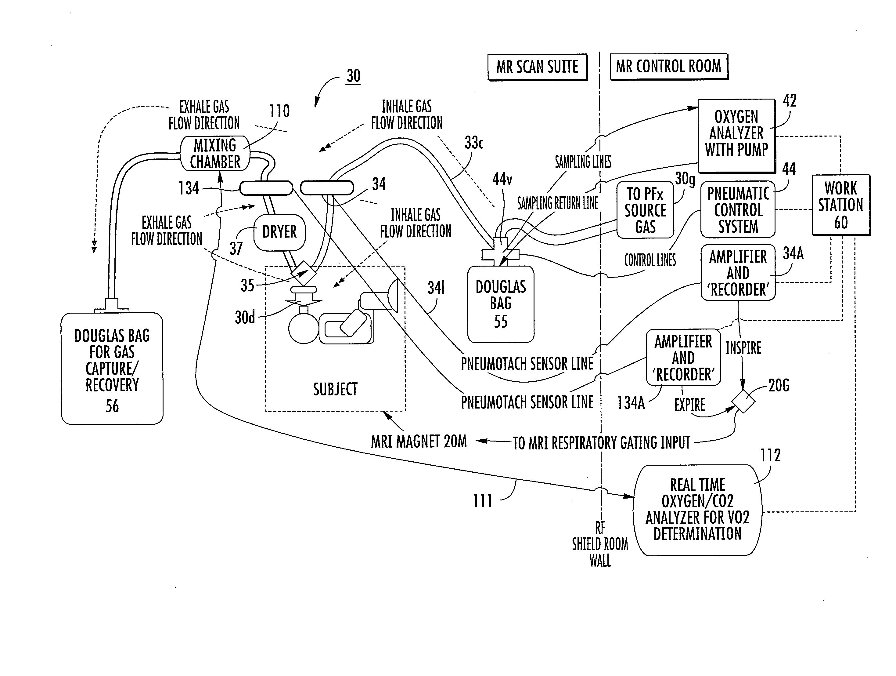
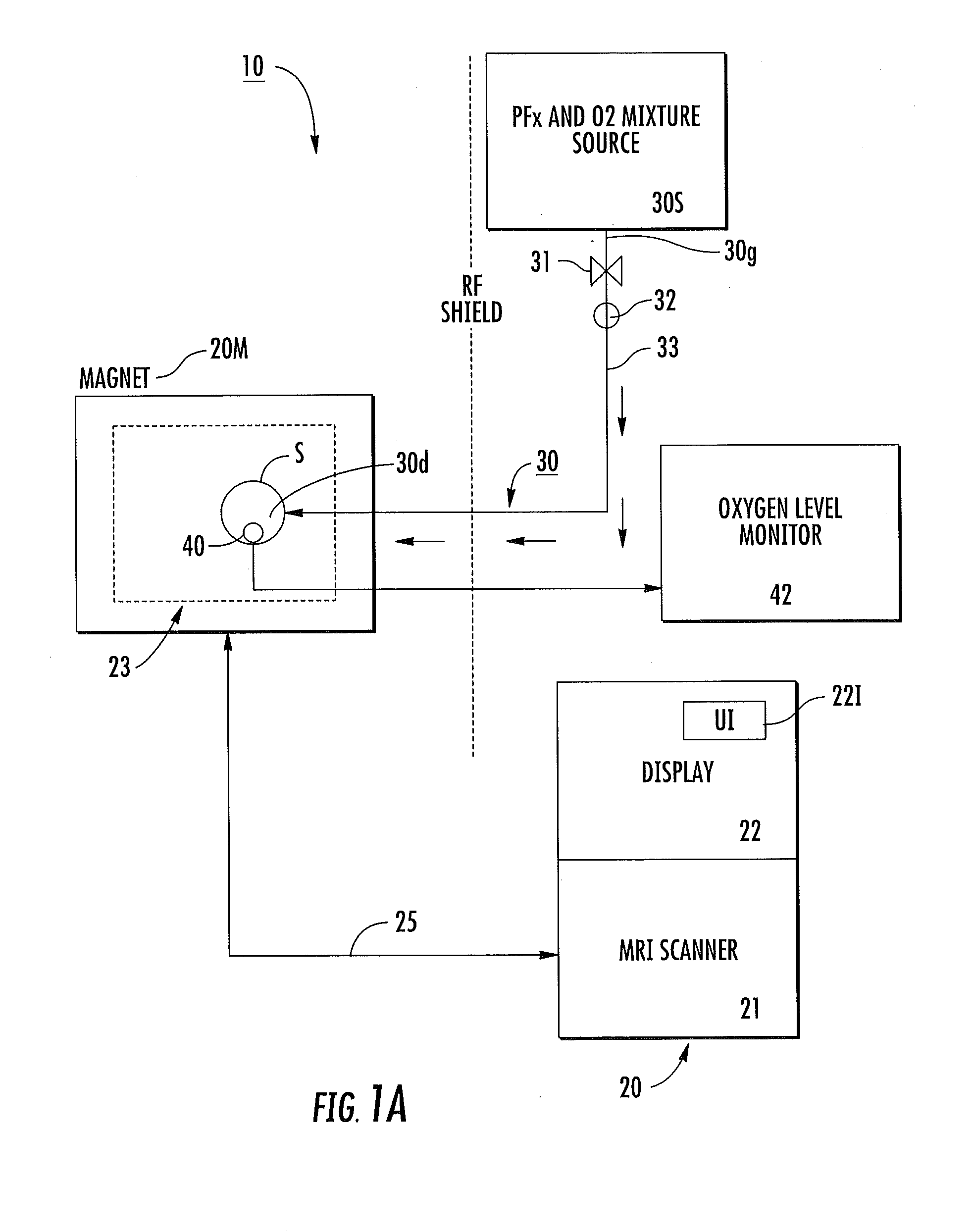
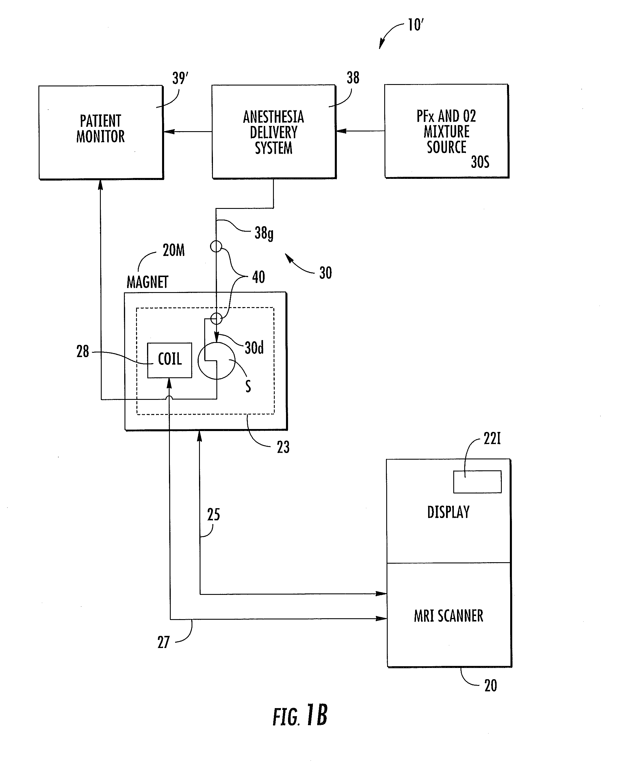
[0065]Still other embodiments are directed to a gas delivery system for an MRI system. The gas delivery systems include: (a) a medical grade gas mixture source of perfluorinated gas and oxygen gas; (b) a mouthpiece and / or mask residing downstream of the gas mixture source configured to reside inside a magnetic field of the MRI system; and (c) an inspire flow path extending from the gas mixture source to the mouthpiece and / or mask. The flow path can include: a first Douglas bag in fluid communication with the gas mixture source; and a first spirometer filter residing downstream of the first Douglas bag. The gas delivery system can be a passive system that allows a patient to “breath-hold” and freely breathe the gas mixture.

Problems solved by technology

There is clear recognition that COPD includes both emphysema and small airway disease; however, there is little appreciation of how to identify COPD early—before there is significant airflow obstruction and clinical impairment.
While spirometry is low cost and widely available, it does not yield any regional information about ventilation distribution or ventilation dynamics in the lung.
Also, these modalities deliver ionizing radiation, which limits their repeat use in patients, especially in clinical trials.
Unfortunately, this technology is relatively expensive and has not been widely disseminated.

Method used


Image

  • Systems, methods, compositions and devices for in vivo magnetic resonance imaging of lungs using perfluorinated gas mixtures
  • Systems, methods, compositions and devices for in vivo magnetic resonance imaging of lungs using perfluorinated gas mixtures
  • Systems, methods, compositions and devices for in vivo magnetic resonance imaging of lungs using perfluorinated gas mixtures

Examples


example 1

Breath-Hold Imaging

[0212]FIGS. 8 and 9 illustrate 3D in-vivo human lung images of a healthy 60 year old male obtained using MRI of a PFx / oxygen mixture. It is believed that this imaging methodology has the potential to be a ‘game-changing’ approach to evaluation and understanding of regional human lung ventilation as well as in the efficacy evaluation of pharmaceuticals involved in the treatment of lung disease. FIG. 8 is a panel of images obtained using a single breath hold of PFP. FIG. 8 shows 3D slice partitions of the 19F PFx images (scan time 15 seconds). Note the signal in the gas mixture delivery tube in the top frames of FIG. 8. It is contemplated that this signal data can be used as an external calibration standard for PFx signal.

[0213]FIG. 9 is a panel of three sets of images with the center set being a 3D fusion of 1H images (which are shown on the far left set of images as “base” images) co-registered with the PFx images (shown as the set on images on the far right). The...

example 2

MRI Cine Images of Lung Function

[0218]Cine images of breathing lungs can be generated as a “movie of breathing” that shows lung air spaces as they fill and evacuate during a respiratory cycle (including inhalation to exhalation) or during a FEV 1 maneuver to visualize or show where the gas goes or stays and / or ventilation defects. The image data can be obtained over a plurality of respiratory cycles and the corresponding images (e.g., image slices) can be co-registered illustrating anatomy changes and ventilation data during the respiratory cycle.

[0219]The cine images can be gated cine images. That is, the respiratory waveform can be monitored and the image data registered (gated) to the part of the cycle during which it was obtained. A typical respiratory cycle (inhale to exhale) is about 2-5 seconds long. Several images can be taken, each in less than one second, and temporally registered or matched to the associated part of the respiratory cycle. The image data can be obtained ov...

example 3

Free-Breathing Scans

[0221]19F gas (and 1H) images of the human lung and airways can be obtained in free-breathing delivery to extract regional lung ventilation information. This imaging may be carried out so that the ventilation defects are shown or identified in near real-time. Particular disease states may make one of the PFx agents preferable over another such as where breathing cycle time for respiratory / gated imaging may be disease dependent.

[0222]The PFx gas agents will allow very rapid imaging with the possibility of near real time imaging of ventilation dynamics.

[0223]The image signal acquisition can be carried out to provide gated / cine images with free breathing strategies. Other cycle-based image generation techniques can also be used, such as, for example, spiral and propeller with passive “free-breathing” delivery of the PFx / O2 gas mixtures and room air to provide dynamic visualizations or cines of lung motion (which can include ventilation information / data for both inha...


PUM


Abstract

Systems and methods for generating MRI images the lungs and / or airways of a subject using a medical grade gas mixture comprises between about 20-79% inert perfluorinated gas and at least about 20.5% oxygen gas. The images are generated using acquired 19F magnetic resonance image (MRI) signal data associated with the perfluorinated gas and oxygen mixture.

Description

RELATED APPLICATIONS[0001]This application claims the benefit of and priority to U.S. Provisional Application Ser. No. 61 / 305,025 filed Feb. 16, 2010 and U.S. Provisional Application Ser. No. 61 / 435,599 filed Jan. 24, 2011, the contents of which are hereby incorporated by reference as if recited in full herein.FIELD OF THE INVENTION[0002]The present invention relates to non-invasive in vivo 19F Magnetic Resonance Imaging (“MRI”) using perfluorinated gas mixtures.BACKGROUND OF THE INVENTION[0003]The Centers for Disease Control (CDC) has stated that COPD (chronic obstructive pulmonary disease) has escalated to the 3rd leading cause of death in this country. See, A M, Miniño, Xu J Q, and Kochanek K D (2010), ‘Deaths: Preliminary Data for 2008.’, National Vital Statistics Reports, 59 (2), John Walsh (President, COPD Foundation) remarked that “It's unacceptable that COPD has gone from the fourth leading cause to the third twelve years sooner than what was originally projected. This wake-...

Claims


Application Information

Patent Timeline
Patent Type & Authority Applications(United States)
IPC IPC(8): A61B5/055A61K49/08A61M16/06G06K9/62A61B5/087
CPCA61B5/055A61M2230/435A61B5/7285G01R33/50G01R33/5601G01R33/56325G01R33/56366A61B5/087A61M16/0054A61M16/12G01R33/5673A61M2016/1025A61M2202/0476A61M2230/432A61B5/08A61B5/082A61M16/1055A61M16/107A61M16/085A61B5/0033A61B5/0813A61B2560/0223A61B2576/02
Inventor CHARLES, HAL CECILSOHER, BRIAN J.
Owner DUKE UNIV
Why Eureka
Try Eureka