Cell analyzer

a cell analyzer and analyzer technology, applied in the field of cell analyzers, can solve the problems of large apparatus size, sample cannot be directly observed, damaged cells, etc., and achieve the effect of accurate analysis of gene information and expression information

Inactive Publication Date: 2013-01-31
KANAGAWA INST OF IND SCI & TECH +1
View PDF11 Cites 11 Cited by
  • Summary
  • Abstract
  • Description
  • Claims
  • Application Information

AI Technical Summary

Benefits of technology

[0056]According to the present invention, a minute amount of target cell in blood can be purified at a single cell level to realize accurate analyses of gene information and expression information of the target cell.
[0057]According to the present invention, whether subject cells are clustered or not (whether it is in the form of an isolated single cell or not) can be identified.
[0058]According to the present invention, whether or not apoptosis is induced in a cell can be determined.
[0059]According to the present invention, only the target cell can be separated / purified and collected in real time.
[0060]According to the present invention, only the collected cells can be subjected to determination of the cellular state of each single cell to perform genomic analysis and expression analysis for each single cell.
[0061]According to the present invention, only the collected cell can be recultivated.

Problems solved by technology

However, there are problems with this technique such as that it is expensive, the size of the apparatus is large, an electrical field as high as a few thousand volts is required, a large amount of sample that has been concentrated to a certain degree is required, the cells may be damaged during generation of the droplets, and the sample cannot directly be observed.
In a cell sorter produced by this micro manufacturing technique, however, the response speed for the separation of the sample is slow with respect to observation means.
In addition, there are problems that the concentration of the cells in the sample solution under use has to be increased to a certain degree in advance or otherwise a subtle cell concentration prevents the separation efficiency of the device from being sufficiently enhanced; and further that when a small amount of sample is to be concentrated in a different apparatus, it is not only difficult to collect the concentrated solution without loss but also the cells might be contaminated during this cumbersome pretreatment stage, which are unfavorable in regenerative medicine and the like.
Currently, although detection of cancerous tissues has been tremendously improved owing to the improvement in MRI (magnetic resonance imaging) and CT (computed tomography), no technique that is superior to the assessment method by biopsy for identifying benign and malignant tumors is present.
There is one issue known for malignant tumors that the cancer cells metastasize from their tissues to other organs due to their ability to invade the blood vessels or the lymph vessels.

Method used


Image

  • Cell analyzer
  • Cell analyzer
  • Cell analyzer

Examples


Embodiment Construction

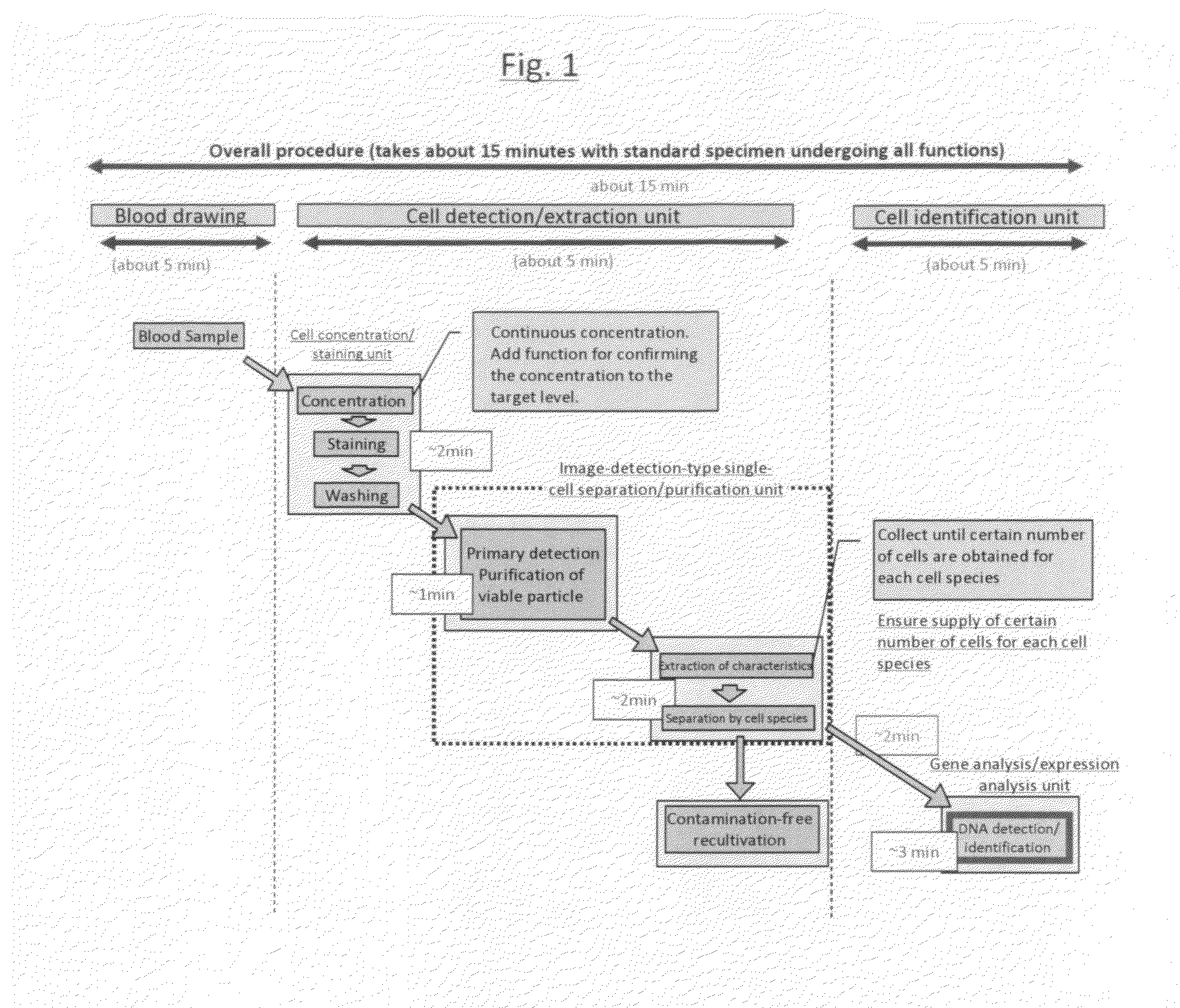
[0083]A cell analyzer of the present invention generally comprises:

(1) a cell concentration / staining / decoloring unit that sequentially performs a process including concentration, staining with a fluorescence antibody label (or in the case where recultivation is to be performed, if necessary, a reversible fluorescence-labeled marker such as aptamer) and washing of cells;

(2) an image-detection-type single-cell separation / purification (cell sorter) unit that captures image data of cell images at about 10,000 images per second from the cells flowing through a micro flow-path formed on a chip substrate, and purifies 10,000 cells per second in real time based on the analysis results of the image information;

(3) a single-cell genomic analysis / expression analysis unit that determines the cellular state at a single cell level;

(4) a transport unit for transporting a sample solution among each of the above-mentioned units; and

(5) a control / analysis unit for controlling performance of each of t...


PUM

PropertyMeasurementUnit
temperaturesaaaaaaaaaa
external forceaaaaaaaaaa

Abstract

Provided is a cell concentration and purification device, having: a function of continuously concentrating cells; a function of then subsequently disposing the cells continuously in a specific region of a channel; a function of simultaneously recognizing, based on an image, the shape and fluorescence emission of each single cell; and a function of recognizing the cells and then separating and purifying the same based on the data relating to the shape and fluorescence emission thereof.

Description

TECHNICAL FIELD[0001]The present invention relates to a cell analyzer.BACKGROUND ART[0002]In biological tissues of a multicellular organism, various cells play their own roles and maintain harmonious functions as a whole. Alternatively, a part of the cells turn into cancer (herein, “cancer” and “tumor” are collectively referred to as “cancer”), then it becomes a neoplasm that is different from the surrounding region. There is not necessarily a clear-cut boundary between the cancerous region and a normal tissue region which is remote from the cancerous region, and the regions surrounding the cancerous region are also affected to some extent. Therefore, in order to analyze a function of cells in an organ tissue, it is necessary to separate and analyze a small number of cells present in a small region as simple as possible in a short period of time and with minimum loss.[0003]In addition, in the field of regenerative medicine, organ stem cells are separated from a tissue and recultivat...

Claims


Application Information

Patent Timeline
Patent Type & Authority Applications(United States)
IPC IPC(8): C12M1/42
CPCG01N1/31G01N1/4077G01N15/1459G01N15/1463G01N21/6408C12N1/066G01N2001/4094G01N2015/149G01N2021/6482G01N1/34G01N21/6428Y10T436/11
Inventor TERAZONO, HIDEYUKIYASUDA, KENJIHAYASHI, MASAHITOTAKEI, HIROYUKIHATTORI, AKIHIRO
Owner KANAGAWA INST OF IND SCI & TECH
Why Eureka
Try Eureka