Use of a Proteolytic Enzyme for the Modification of the Cell Surface of a Stem Cell

a technology of stem cell and proteolytic enzyme, which is applied in the field of stem cell cell cell cell cell cell surface modification by proteolytic enzyme, can solve the problems of unwanted transplantation of cells and higher clinical complications, and achieve the effect of hindering and/or preventing the transition of stem cells

Inactive Publication Date: 2013-02-07
GLYKOS FINLAND
View PDF1 Cites 0 Cited by
  • Summary
  • Abstract
  • Description
  • Claims
  • Application Information

AI Technical Summary

Benefits of technology

[0014]In particular, an object of the present invention is to provide a method of assisting therapeutic stem cells to the target organ(s) or tissues. Another object of the present invention is to provide a method of hindering and / or preventing the transition of stem cells from blood stream to organs which are not the actual target ones, e.g., the lungs and / or liver. A further object of the present invention is to provide a method of modifying and / or altering the distribution behaviour of cells used for cellular therapy.

Problems solved by technology

A problem related to stem cell transplantation as done using current standards is entrapment (may also be called as “distribution” or “homing”) of the transplanted cells to unwanted organs or tissues.
However, by this approach the increased dose unfortunately tends to result in higher rates of clinical complications, for example, graft-versus-host disease, a life threatening condition after HSC transplantation.

Method used


Image

  • Use of a Proteolytic Enzyme for the Modification of the Cell Surface of a Stem Cell
  • Use of a Proteolytic Enzyme for the Modification of the Cell Surface of a Stem Cell
  • Use of a Proteolytic Enzyme for the Modification of the Cell Surface of a Stem Cell

Examples


example 1

Materials and Methods

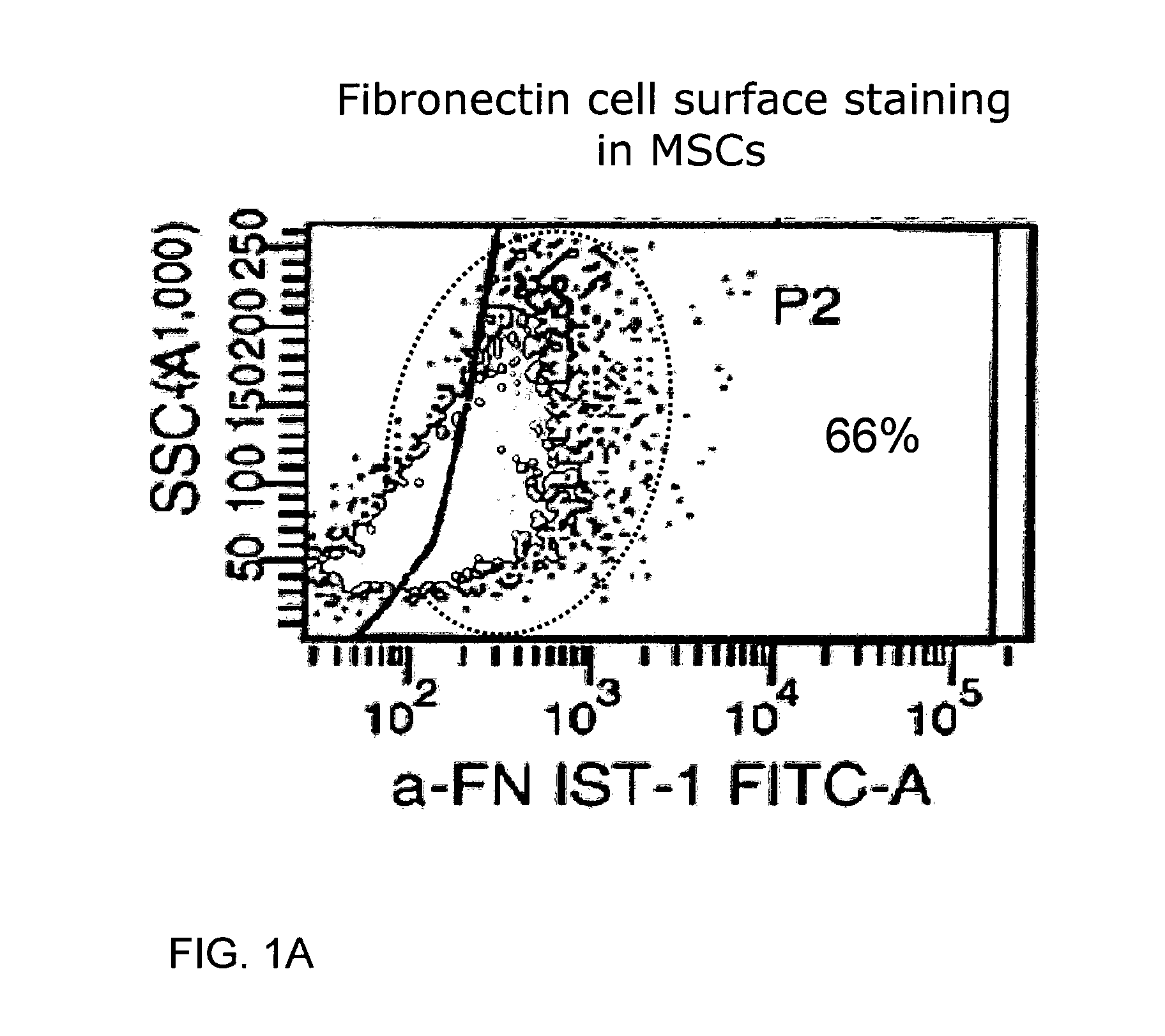
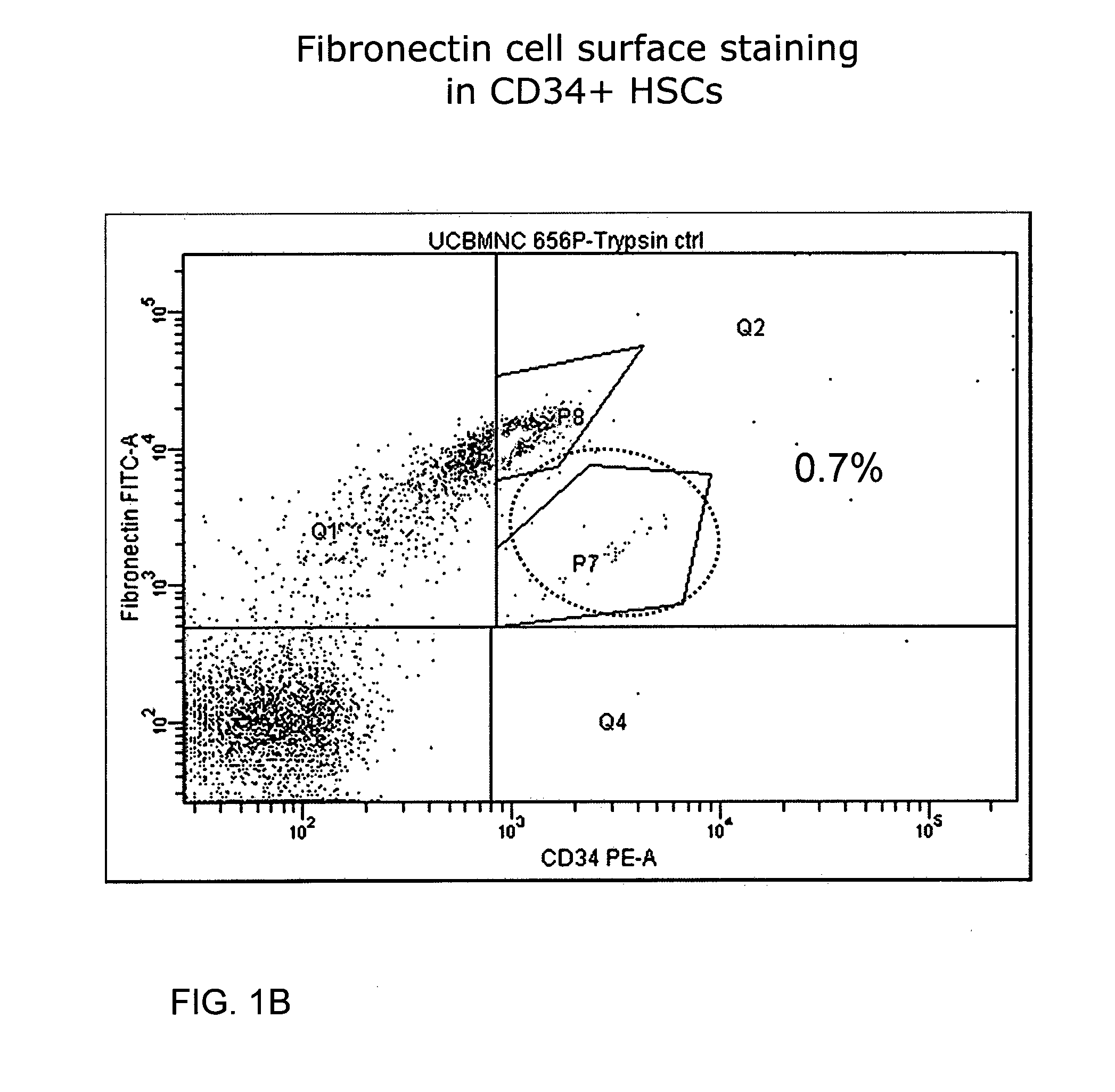
[0065]Umbilical cord blood-derived mesenchymal stem cell (UCBMSC) 594P in p2, UCBMSC 454T(7) in p4 and Ficoll-isolated UCB-derived mononuclear cells (MNCs) were used in the experiments. The adherent MSCs were detached in 70-100% confluency with trypsin (TryPLe Express, Invitrogen) and the trypsinization was stopped within 4 minutes with excess culture media. The cells were labelled for flow cytometric analysis with 2 μl of the anti-fibronectin antibody (#ab6327, abcam, FIG. 17) and PE-conjugated anti-CD34 antibody (#130-081-002, Miltenyi Biotec) per 1×10e5 cells in PBS w / Ca & Mg pH 7.2+0.5% bovine serum albumin (BSA) for 30 minutes on ice. After washing with excess labelling buffer, secondary antibody staining was done with Alexa 488-conjugated goat-anti mouse IgG (H+L) diluted 1:500. The labelled cells were run with a FACSAria (BD) flow cytometer and the results were analyzed with the FACSDiva software (BD).

Results

[0066]MSCs: The UCBMSCs were concluded to be c...

example 2

Materials and Methods

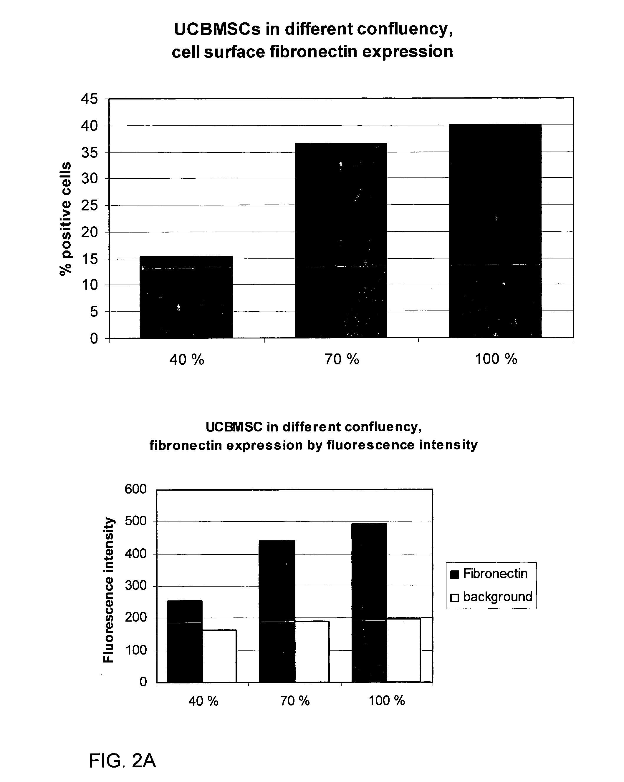
[0069]Confluency Experiments:

[0070]Umbilical cord blood-derived mesenchymal stem cell (UCBMSC) 454T(7) in p6 were used in the confluency experiments. The cells were plated in different densities to yield different levels of confluency on the same analysis day. The MSCs were analyzed at 40%, 70% and 100% confluency and were simultaneously detached with trypsin (TryPLe Express, Invitrogen). The trypsinization was stopped within 4 minutes with excess culture media. The cells were labelled for flow cytometric analysis with 2 μl of the anti-fibronectin antibody (#ab6327, abcam) per 1×10e5 cells in PBS w / Ca & Mg pH 7.2+0.5% bovine serum albumin (BSA; ultrapure, Sigma) for 30 minutes on ice. After washing with excess labelling buffer, secondary antibody staining was done with Alexa 488-conjugated goat-anti mouse IgG (H+L) diluted 1:500. The labelled cells were run with a FACSAria (BD) flow cytometer and the results were analyzed with the FACSDiva software (BD).

[0071]Su...

example 3

Materials and Methods

[0074]Pediatric human bone marrow-derived mesenchymal stem cell (BMMSC) line M2 in passage 6 was used for the study. The subconfluent cells were detached with either trypsin (TryPLE Express, Invitrogen) or 0.5% pronase in PBS-0.5 mM EDTA. Detachment was stopped after 4 minutes by adding excess culture media. Viability was determined by trypan blue exclusion. The mitochondrial inner potential was measured with the JC-1label (Molecular Probes, Invitrogen) and flow cytometry.

Results

[0075]As compared to trypsin, the pronase detachment protocol (maximum concentration tested 0.5% pronase in PBS-EDTA) was as fast as the trypsin detachment protocol. Pronase detachment produced very viable, one-cell MSC suspensions without any cell aggregates. The cells had >95% viability (equal to trypsin) as determined by Trypan blue exclusion. Pronase-detached cells exhibited a better mitochondrial inner potential as studied with the JC-1 label as compared to trypsinized cells (see F...


PUM


Abstract

The present invention relates to a stem cell and/or a population thereof having a specific profile of cell surface proteins and/or proteoglycans. The present invention also relates to use of a proteolytic enzyme in the modification of the cell surface of a stem cell. The present invention further relates to a method of modifying the cell surface of a stem cell by treatment with a proteolytic enzyme.

Description

FIELD OF THE INVENTION[0001]The present invention relates to a stem cell and / or a population thereof having a specific profile of cell surface proteins and / or proteoglycans. The present invention also relates to use of a proteolytic enzyme in the modification of the cell surface of a stem cell. The present invention further relates to a method of modifying the cell surface of a stem cell by treatment with a proteolytic enzyme.BACKGROUND OF THE INVENTION[0002]Proteolytic enzymes are a large group of enzymes which are involved in digesting protein chains into shorter fragments by splitting the peptide bonds that link amino acid residues together. Some of them are able to detach the terminal amino acids from the peptide or protein chain (exopeptidases, such as aminopeptidases) and the others attack internal peptide bonds of a protein (endopeptidases, such as trypsin, chymotrypsin, pepsin, papain, elastase). Proteolytic enzymes can be divided into four major groups according to the char...

Claims


Application Information

Patent Timeline
Patent Type & Authority Applications(United States)
IPC IPC(8): C12N5/0789
CPCA61K35/12C12N5/0006C12N2509/00C12N5/0665C12N2501/734C12N5/0663
Inventor NYSTEDT, JOHANNAHAKKARAINEN, TANJALEHENKARI, PETRIKERKELÄ, ERJAVALMU, LEENA
Owner GLYKOS FINLAND
Why Eureka
Try Eureka