
Drug Delivery Apparatus

a technology of delivery device and ear, which is applied in the direction of suction device, catheter, other medical devices, etc., can solve the problems of not being able to reduce not being able to address hearing loss prevention, and not being able to increase the basic capacity of a damaged ear, so as to reduce the side effects of implant surgery and improve the performance of the prosthesis

Active Publication Date: 2013-02-28
MASSACHUSETTS EYE & EAR INFARY +1
View PDF8 Cites 18 Cited by
  • Summary
  • Abstract
  • Description
  • Claims
  • Application Information

AI Technical Summary

Benefits of technology

This patent is for an apparatus that can deliver drugs to the cochlea to treat hearing disorders, reduce side effects of implant surgery, and improve performance of a cochlear prosthesis. The apparatus includes a pressurized drug supply reservoir and a sensor to measure properties of an endogenous fluid that enters the apparatus. The apparatus can be integrated with a cochlear implant or other auditory system for electrical stimulation. The technical effect of this invention is the ability to deliver targeted drugs to the cochlea for improved treatment of hearing disorders.

Problems solved by technology

However, such treatments cannot address hearing loss prevention, cannot minimize hearing loss progression and, even with optimal device fitting, cannot increase a damaged ear's basic capacity.
As a result, many users continue to experience significant communication difficulties.
The crux of the problem in SNHL is loss of the delicate cochlear sensory cells that detect the exquisitely small mechanical vibrations associated with sound.
In human ears, once lost or damaged, these sensory cells do not regenerate and this compromise is often followed by secondary degeneration of auditory neurons.
This ‘protection’, however, also prevents certain molecules with potentially therapeutic effect from gaining access to their inner ear targets.
Current otologic practice requires drug delivery to the inner ear, but uses inefficient routes.
Their clinical usefulness, however, is limited by undesirable side effects arising from the high systemic doses required to achieve sufficient cochlear fluid levels of drug to produce the intended inner ear effects.
Delivery of drugs to the middle ear reduces systemic side effects, but access to the inner ear is unpredictable.
A major limitation of these methods, however, is the inability to precisely control the amount of drug that diffuses from the middle ear through the RWM into the inner ear.
This becomes even more problematic when considering delivery of coplex macromolecules with limited diffusion coefficients and those requiring sequenced delivery.
Additionally, the bolus application used by certain existing systems makes them poorly suited for direct inner ear delivery.
Although such devices may be useful for delivery of low molecular weight, stable, lipid-soluble compounds like steroids, they would not be suitable for the delivery of the unstable macromolecules that ultimately will be the therapeutic compounds with greatest potential benefit.
However, as mentioned above, the delivery of therapeutic agents in a controlled and effective manner is considerably more difficult with respect to tissue structures of the inner ear (e.g. those portions of the ear surrounded by the otic capsule bone and contained within the temporal bone, which is the most dense bone tissue in the entire human body).
The inner ear tissues listed above are of minimal size and only readily accessible through invasive microsurgical procedures.
Imbalances in the pressure and volume levels of such fluids can cause various problems, including but not limited to conditions known as endolymphatic hydrops, endolymphatic hypertension, perilymphatic hypertension, perilymphatic hydrops, perilymphatic fistula, intracochlear fistula, Meniere's disease, tinnitus, vertigo, hearing loss related to hair cell or ganglion cell damage / malfunction, and ruptures in various membrane structures within the ear.
For example, it has been determined that dry compounds, which may be more stable than aqueous ones, cannot be used in a conventional infuser.
Moreover, in the application to inner ear diseases, dosage to the relevant tissues of the cochlea can be difficult or impossible to assess and control by the methods described above, and no device has been provided for programmable long-term delivery, either to the middle ear or inner ear.
Known methods require a relatively complicated mechanism to achieve mixing and circulating flow between reservoir and patient.
These more complicated methods include having two tubes entering the patient, rather than just one, or having a two-way pump, two pumps, or a switching valve at the pump.
Notably, existing drug-delivery technology is typically not appropriate for long-term programmable infusion into the inner ear.
These systems range from passive devices, which have a low level of predictability in their dispense rates, to electronically-controlled rate dispensers, and finally to fully programmable infusers.
They are generally short to medium term delivery devices (less than six weeks) with unalterable, non-constant delivery profiles.
However, because they use macro scale conventionally fabricated pumps, these systems are relatively large.
This exemplary method may be effective for some applications, but is generally not effective when clinical requirements necessitate specific flow conditions, when a particular form of drug storage is desired, or when power conservation is a major factor in system design.

Method used


Image

  • Drug Delivery Apparatus
  • Drug Delivery Apparatus
  • Drug Delivery Apparatus

Examples


Embodiment Construction

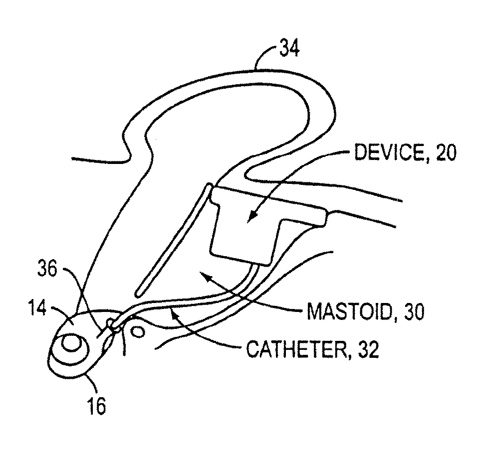
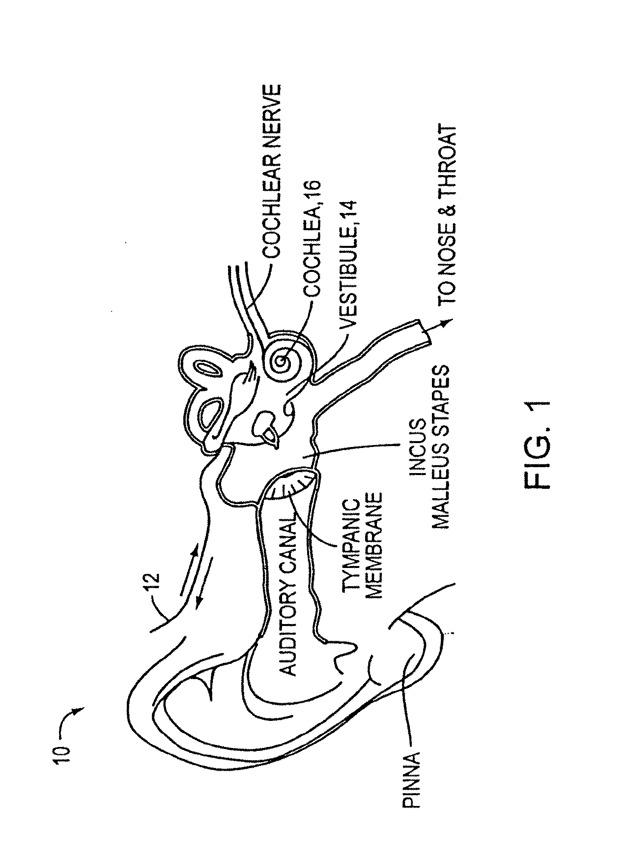
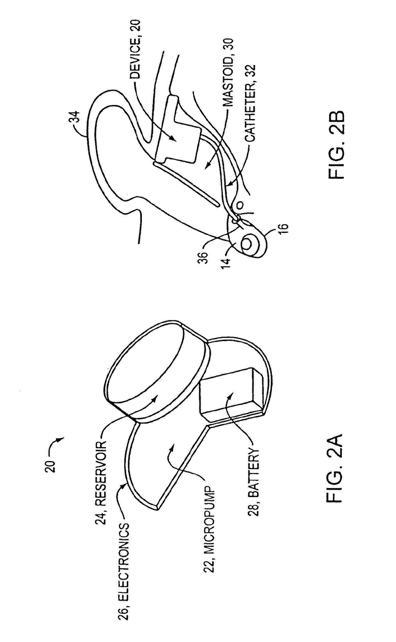
[0056]As discussed above, conventional drug infusers utilize macroscale machined components to pump liquid drugs from a reservoir. Various embodiments of the present invention replace these components with a synthesis of micropumps and MEMS solutions for drug storage and release, which results in smaller devices with greater functionality. This opens up the inner ear and other previously inaccessible locations in the body to new direct treatment, without the side effects of systemic delivery.

[0057]Microfluidics and microelectromechanical systems (MEMS) capability can be used for drug delivery applications, to allow or provide a controlled rate, low drug volume, and / or liquid formulation (e.g., for an implantable inner ear delivery system). In an example embodiment, a fluidic system having a closed loop microfluidic flow controller can be used with animal test apparatus. In one embodiment of the current invention, an implanted recirculating delivery system can be used in therapy for ...


PUM


Abstract

An implantable drug delivery apparatus for delivering a drug into a bodily fluid in a bodily cavity of a patient over a period of time includes a drug supply reservoir to supply drug into a delivery channel and an actuator for delivering the drug to a predetermined location in the bodily cavity of the patient, such as, for example, a cochlea of a human ear. The drug is loaded into the delivery channel while producing substantially negligible flow at an outlet of the delivery channel.

Description

CROSS-REFERENCE TO RELATED APPLICATION[0001]This application claims priority to and the benefit of U.S. Provisional Patent Application No. 61 / 438,934, filed Feb. 2, 2011, which is incorporated herein by reference in its entirety.STATEMENT REGARDING FEDERALLY SPONSORED RESEARCH OR DEVELOPMENT[0002]This invention was made with government support under Grant No. 2 R01 DC006848-04A1 awarded by the National Institutes of Health. The government has certain rights in the invention.FIELD OF THE INVENTION[0003]The present invention relates generally to the field of drug delivery devices employing catheters and / or cannulas to transport fluid from a reservoir to a patient and, more particularly, to a device for introducing a drug into a patient's bodily fluid, such as, for example, into perilymph in the human ear, as well as to methods for infusing drugs into cochlea for treatment of hearing loss and other disorders of hearing and vestibular function.BACKGROUND OF THE INVENTION[0004]Sensorineu...

Claims


Application Information

Patent Timeline
IPC IPC(8): A61M31/00
CPCA61M2210/0668A61M5/14276A61M31/002
Inventor FIERING, JASON O.MESCHER, MARK J.PARARAS, ERIN E.BORENSTEIN, JEFFREY T.SEWELL, WILLIAM F.KUJAWA, SHARON G.MCKENNA, MICHAEL J.KIM, ERNEST S.
Owner MASSACHUSETTS EYE & EAR INFARY
Learn More