Renal nerve denervation via the renal pelvis

Active Publication Date: 2013-07-11
VERVE MEDICAL
View PDF5 Cites 22 Cited by
  • Summary
  • Abstract
  • Description
  • Claims
  • Application Information

AI Technical Summary

Benefits of technology

[0010]The wall of the renal pelvis is a particularly rich source for afferent sensory nerves which are found in the urothelium which lies immediately adjacent to the renal pelvis. They are also found in rich supply in the intermediate submucosale layer which is closest to the urothelium. The renal pelvis wall is also a source for efferent nerves which are found in both the intermediate and outer submucosale layers. Thus, the treatments of the present invention which exchange energy or deliver active agents from the renal pelvis may be particularly effective in treating the afferent sensory nerves which are presently believed to be principally responsible for the reduction of hypertension.
[0011]The present invention relies on introducing or advancing the effector into the interior of the renal pelvis by a minimally invasive approach or access. Usually the access will be through the urinary tract and thus not require percutaneous penetration (and thus may be performed as a “natural orifice surgery”). Alternatively, the access could be achieved through known laproscopic or other percutaneous techniques relying on access penetrations through the abdominal wall and advancement of tools through the body of the kidney in order to access

Problems solved by technology

Such laparoscopic techniques are on the one hand disadvantageous because they require such tissue penetrations but on the other hand are advantageous in that they

Method used


Image

  • Renal nerve denervation via the renal pelvis
  • Renal nerve denervation via the renal pelvis
  • Renal nerve denervation via the renal pelvis

Examples


Embodiment Construction

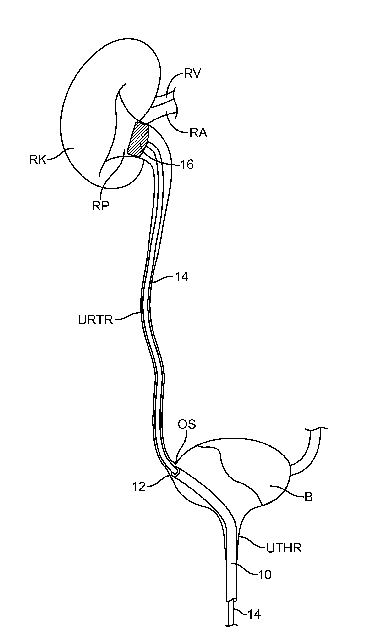
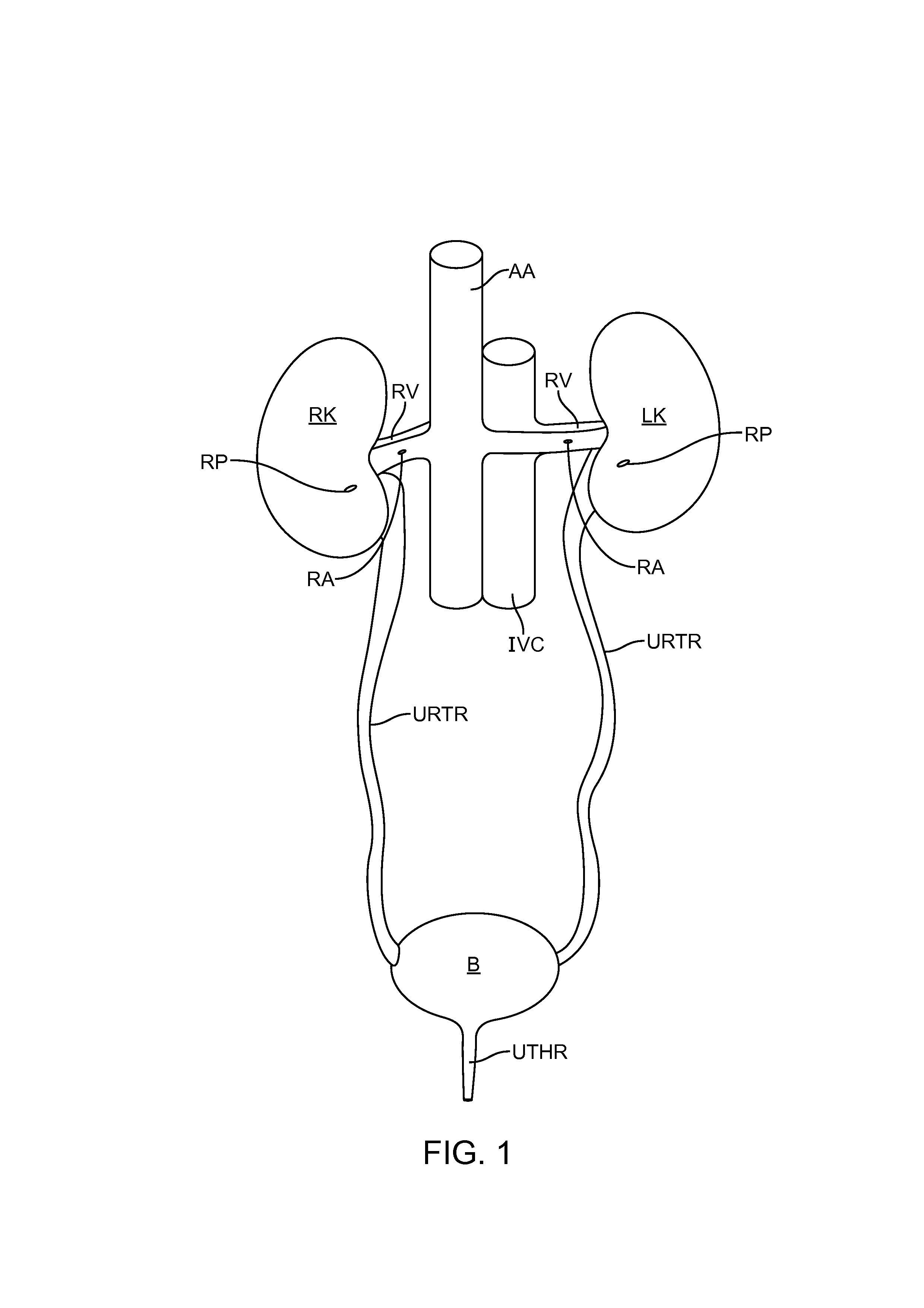
[0039]A patient's urinary tract is diagrammatically illustrated in FIG. 1. The urinary tract includes the bladder B, which receives urine from the right and left kidneys RK and LK and drains the urine through the urethra UTHR. The kidneys each receive oxygenated blood through the renal artery RA from the abdominal aorta AA and blood from the kidneys is returned through the renal vein RV to the inferior vena cava IVC. Of particular interest to the present invention, the urine which is processed in the kidney is received in an interior cavity of each kidney referred to as the renal pelvis RP which acts as a funnel and delivers the urine into the top of the ureter URTR. The methods and protocols of the present invention will be performed within the interior of the renal pelvis RP in order to treat the renal nerves within the walls of the renal pelvis as well as the nerves surrounding the renal arteries within the adventitia and adipose tissue and to a lesser extent surrounding the rena...


PUM


Abstract

Apparatus, systems, and methods provide access to the renal pelvis of a kidney to treat renal nerves embedded in tissue surrounding the renal pelvis. Access to the renal pelvis may be via the urinary tract or via minimally invasive incisions through the abdomen and kidney tissue. Treatment is effected by exchanging energy, typically delivering heat or extracting heat through a wall of the renal pelvis, or by delivering active substances.

Description

CROSS-REFERENCES TO RELATED APPLICATIONS[0001]The present application claims the benefit of U.S. Provisional Patent Application No. 61 / 506,976 (Attorney Docket No. 42532-703.101), filed Jul. 12, 2011, the entire contents of which are incorporated herein by reference.BACKGROUND OF THE INVENTION[0002]1. Field of the invention[0003]The present invention relates generally to medical devices, systems, apparatus, and methods for modifying nerve function and treating disease. More particularly, the present invention relates to methods and apparatus for exchanging energy or delivering active agents through the renal pelvis to modify sympathetic nerve activity in the adventitia of arteries and / or veins that surround the external surface of the renal pelvis in the kidney and in the afferent and efferent nerves within the muscles layers, urothelium and submucosa of the renal pelvis.[0004]Hypertension, or high blood pressure, is a significant and growing health risk throughout the world. Hypert...

Claims


Application Information

Patent Timeline
IPC IPC(8): A61B18/00A61M25/10
CPCA61B18/00A61M25/10A61N7/022A61B18/08A61B18/1492A61B18/1815A61B2018/0016A61B2018/0022A61B2018/00267A61B2018/00291A61B2018/00434A61B2018/00511A61B2018/044A61B2018/046A61B2018/143A61B2018/1475A61B2018/00404
Inventor BUELNA, TERRENCE J.
Owner VERVE MEDICAL
Why Eureka
Try Eureka