
Steerable catheter navigation with the use of interference ultrasonography

a technology of interference ultrasonography and catheter navigation, which is applied in the field of ultrasonic imaging systems and methods, can solve the problems of inability to detect the motion of the cardiac wall, limited spatial resolution, and the cost of employing this method and the required stereotactic system is rather prohibitive, and achieves the effect of high degree of accuracy and facilitate unique identification of the motion

Inactive Publication Date: 2013-08-08
MAYO FOUND FOR MEDICAL EDUCATION & RES
View PDF2 Cites 12 Cited by
  • Summary
  • Abstract
  • Description
  • Claims
  • Application Information

AI Technical Summary

Benefits of technology

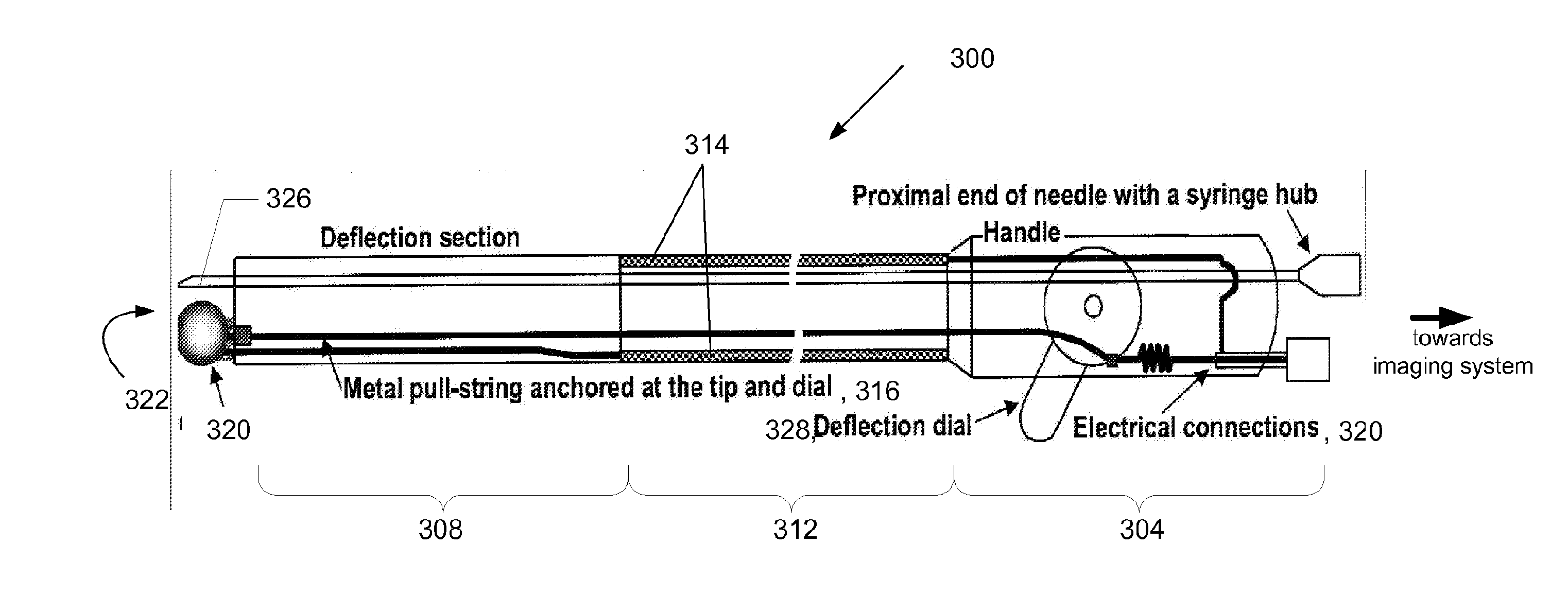
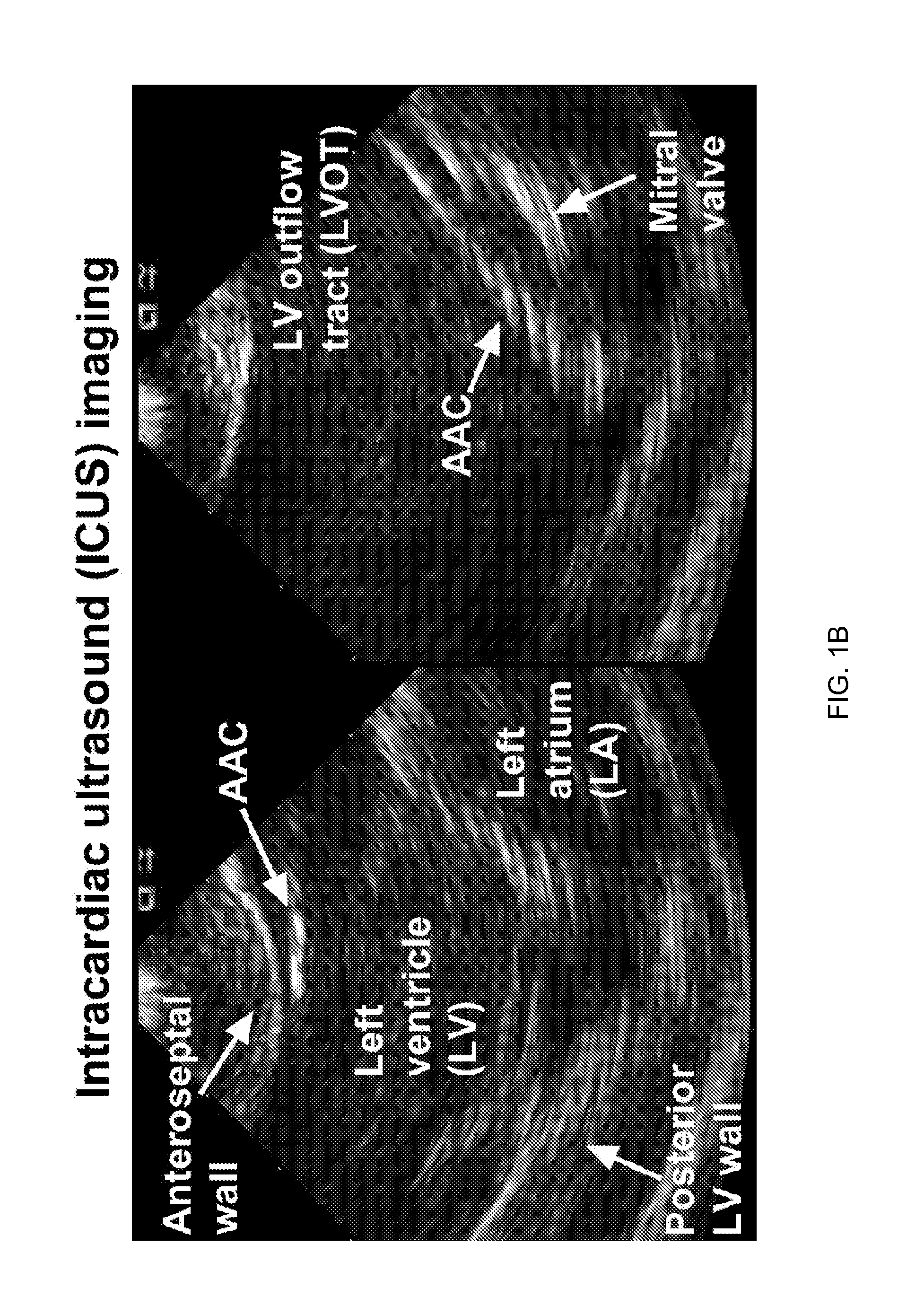
The invention provides an acoustically-active catheter (AAC) that uses interferometric signals to actively track and navigate within a bodily system, such as the cardiovascular system, using ultrasound imaging. The AAC has a unique identified tip that produces acoustic waves that interfere with each other, creating an acoustic interference signal that can be detected and used for accurate tracking and navigation. The system uses color-coding to easily identify the motion of the AAC within the tissue. The AAC can also be used for active navigation in the absence of any conventional interferometric signal, making use of the acoustic interference signal as an informative interferometric signal. The invention provides an improved method for tracking and controlling the catheter's tip in real-time, allowing for better navigation and positioning within the bodily system.

Problems solved by technology

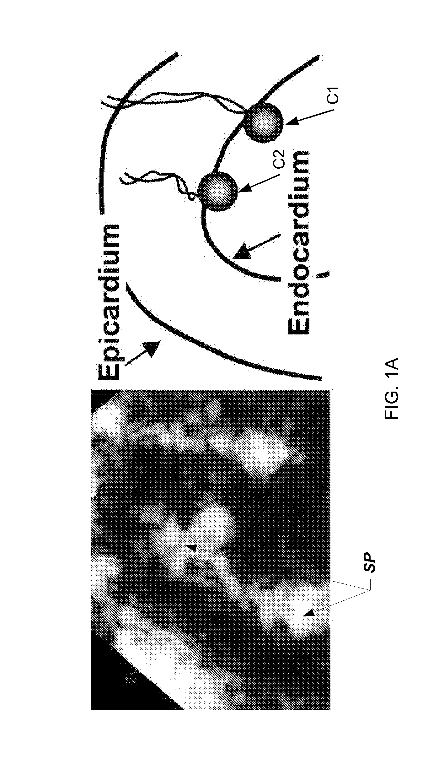
One of such methodologies allows for a sparse electromechanical mapping of the endocardial surface of the left ventricle (LV) by employing a so-called NOGA catheter that is placed into the LV under the X-ray control, which is required because NOGA lacks a capability to provide an anatomical image of the heart.
This approach has a number of drawbacks such as exposure to ionizing radiation for both a patient and medical personnel, projection of large three-dimensional (3D) imaging field (through the entire depth of body) onto a two-dimensional (2D) plane, and the necessity to use specialized procedure rooms.
In addition, while NOGA allows for detection of the endocardial surface, it cannot detect the motion of the cardiac wall and has limited spatial resolution.
Finally, the cost of employing this method and the required stereotactic systems is rather prohibitive.
The deficiencies of this not-yet-proven technique include a need in a costly MRI suite, confinement of the catheterization team in proximity to the magnet, and prohibition on use of any metallic instruments.
While a catheter lends itself to being guided with US during insertion, the obtained US-images suffer from speckle patterns and backscatter pattern ambiguity, easily causing errors in the determination of the position of the catheter tip within the cardiovascular system.
It was observed that the simultaneous operation of the sonomicrometer and electrocardiography (echo) suffers from acoustic interference hindering the clarity of US images used for navigation of the sonomicrometric catheter and causing the users to turn off the sonomicrometry system while acquiring echoes and, therefore, causing not saving the sonomicrometric data during imaging.
A skilled artisan shall realize, therefore, that while commonly-available, real-time ultrasonographic systems may satisfy the requirement of being minimally invasive and can be used alone to guide catheters, they have fundamental limitations.
The use of ultrasound imaging system alone to guide catheters within human body (and, in particular, within the heart as discussed herein), suffers from a problem of differentiating the actual tip of the catheter from a bend coming in or out of the 2D Doppler plane.
But the transition from 2D to 3D only converts the problem of reliably localizing the tip of the catheter in or out of a 2D plane to a problem of determining the tip location in or out of a 3D Doppler space.
The fundamental limitations of ultrasound signal propagation, including refraction, attenuation, rather unpredictable backscatter patterns, and signal drop-outs are sources of imaging artifacts that compromise catheter navigation regardless of spatial dimensionality.
Furthermore, the ultrasound image of the catheter tip is often disguised on the background of an image of soft tissue because the backscatter pattern of the catheter is not unique.
As a result, a position of the catheter tip is often misinterpreted or determined inaccurately if the actual tip is located out of the scan plane.
This could lead to accidental injury or piercing of the cardiac wall.

Method used


Image

  • Steerable catheter navigation with the use of interference ultrasonography
  • Steerable catheter navigation with the use of interference ultrasonography
  • Steerable catheter navigation with the use of interference ultrasonography

Examples


Embodiment Construction

[0041]References throughout this specification to “one embodiment,”“an embodiment,”“a related embodiment,” or similar language mean that a particular feature, structure, or characteristic described in connection with the referred to “embodiment” is included in at least one embodiment of the present invention. Thus, appearances of the phrases “in one embodiment,”“in an embodiment,” and similar language throughout this specification may, but do not necessarily, all refer to the same embodiment. It is to be understood that no portion of disclosure, taken on its own and / or in reference to a figure, is intended to provide a complete description of all features of the invention.

[0042]In addition, in drawings, with reference to which the following disclosure may describe features of the invention, like numbers represent the same or similar elements wherever possible. In the drawings, the depicted structural elements are generally not to scale, and certain components are enlarged relative t...


PUM


Abstract

An acoustically-active catheter (AAC) for use with an ultrasound imaging system and a method for tracking the AAC with respect to anatomic target chosen within a body using the interference ultrasonography. A tip of the AAC is equipped with a crystal transmitting an acoustic wave with parameters similar to those of an acoustic wave generated by a transducer of the imaging system so as to produce acoustic interference the strength of which depends on a position of the AAC tip with respect to at least one Doppler scan plane, formed by the imaging transducer, and a distance from a pulsed-wave Doppler sample that is overlapped with the anatomic target. The ultrasound imaging system detects the interference signal and produces a visual and / or audible interference output that indicates the strength of the acoustic interference. Based on the intensity of the interference output, the user can navigate the AAC tip towards the anatomic target. An anatomic target can be chosen within a cardiovascular system.

Description

CROSS-REFERENCE TO RELATED APPLICATIONS[0001]The present application claims benefit of and priority from the U.S. Provisional Patent Application No. 61 / 375,093 filed on Aug. 19, 2010 and titled “Steerable Catheter Navigation With the Use of Interference Ultrasonography.” The disclosure of the above-mentioned application is incorporated herein by reference in its entirety.STATEMENT REGARDING FEDERALLY SPONSORED RESEARCH OR DEVELOPMENT[0002]This invention was made with government support under Grant No. EB009111 awarded by the National Institute of Health. The government has certain rights in this invention.TECHNICAL FIELD[0003]The present invention relates to ultrasonic imaging systems and methods and, more particularly, to ultrasonic systems and methods capable of locating a position of an object, such as a catheter, within a body by utilizing acoustic interference between an acoustic wave generated by the object and the ultrasound (US) wave generated by the imaging system.BACKGROUN...

Claims


Application Information

Patent Timeline
IPC IPC(8): A61B8/00A61B8/12A61B8/08
CPCA61B8/08A61B8/54A61B8/0883A61B8/12A61B8/4477A61B19/5244A61B2017/00243A61B2019/5276A61B2019/5278A61B2019/5285A61B2019/5429A61M25/0108A61M25/0147A61B8/5207A61B8/488A61B8/461A61B8/4483A61B8/445A61B8/4254A61B8/0891A61B8/0841A61B8/463A61B8/466A61B2090/3782A61B2090/3788A61B34/20A61B2090/378A61B2090/3929
Inventor BELOHLAVEK, MAREKMCMAHON, EILEEN M.
Owner MAYO FOUND FOR MEDICAL EDUCATION & RES
Learn More