
Treatment Of Penetrative Injury To The Brain
a brain injury and penetrative technology, applied in the field of brain injury treatment, can solve the problems of clinical trials of these drugs in stroke and traumatic brain injury, and achieve the effect of improving the safety and efficacy of the drug
- Summary
- Abstract
- Description
- Claims
- Application Information
AI Technical Summary
Benefits of technology
Problems solved by technology
Method used
Image

Examples
examples
1. Rat Model of Penetrative Brain Injury
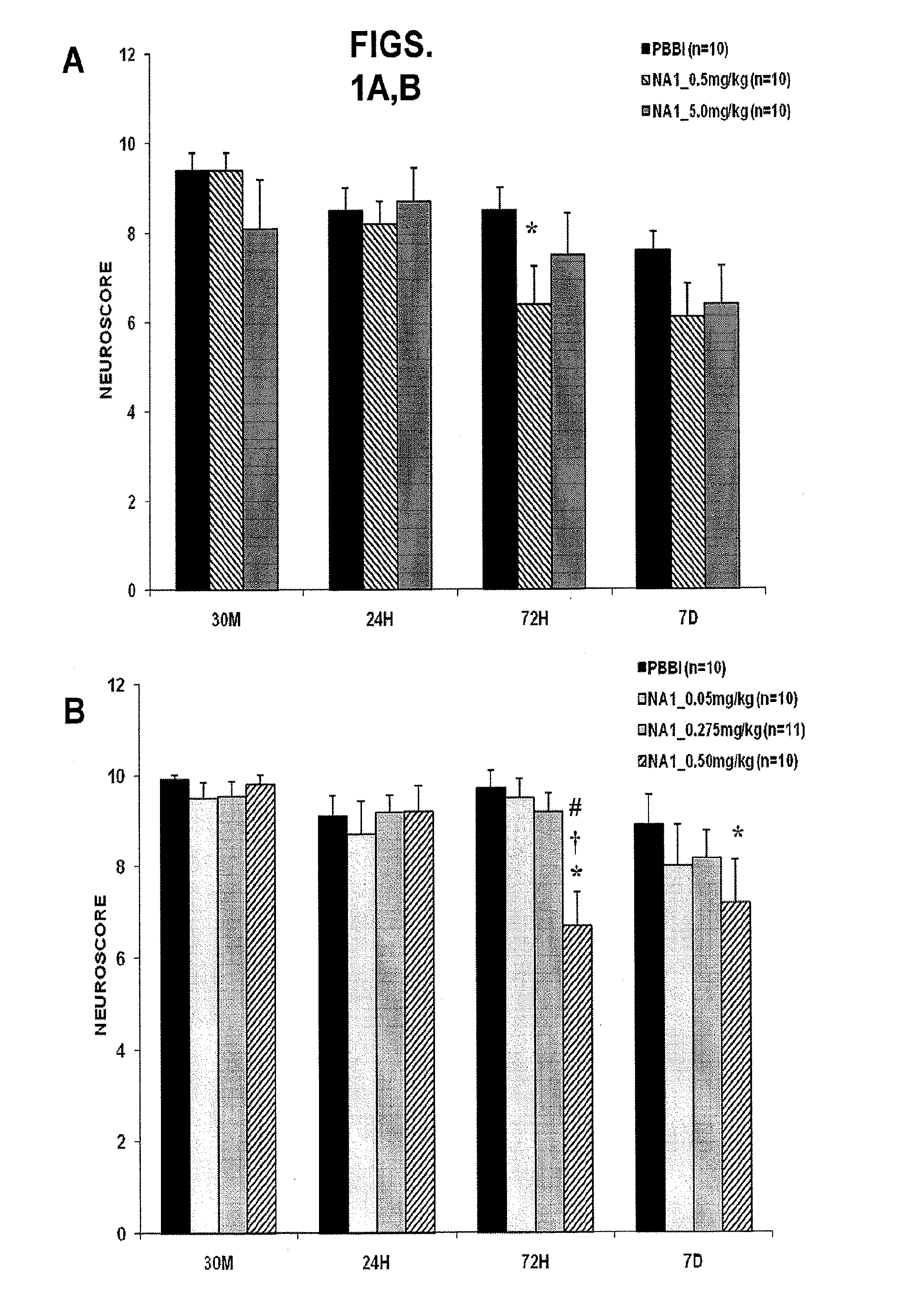
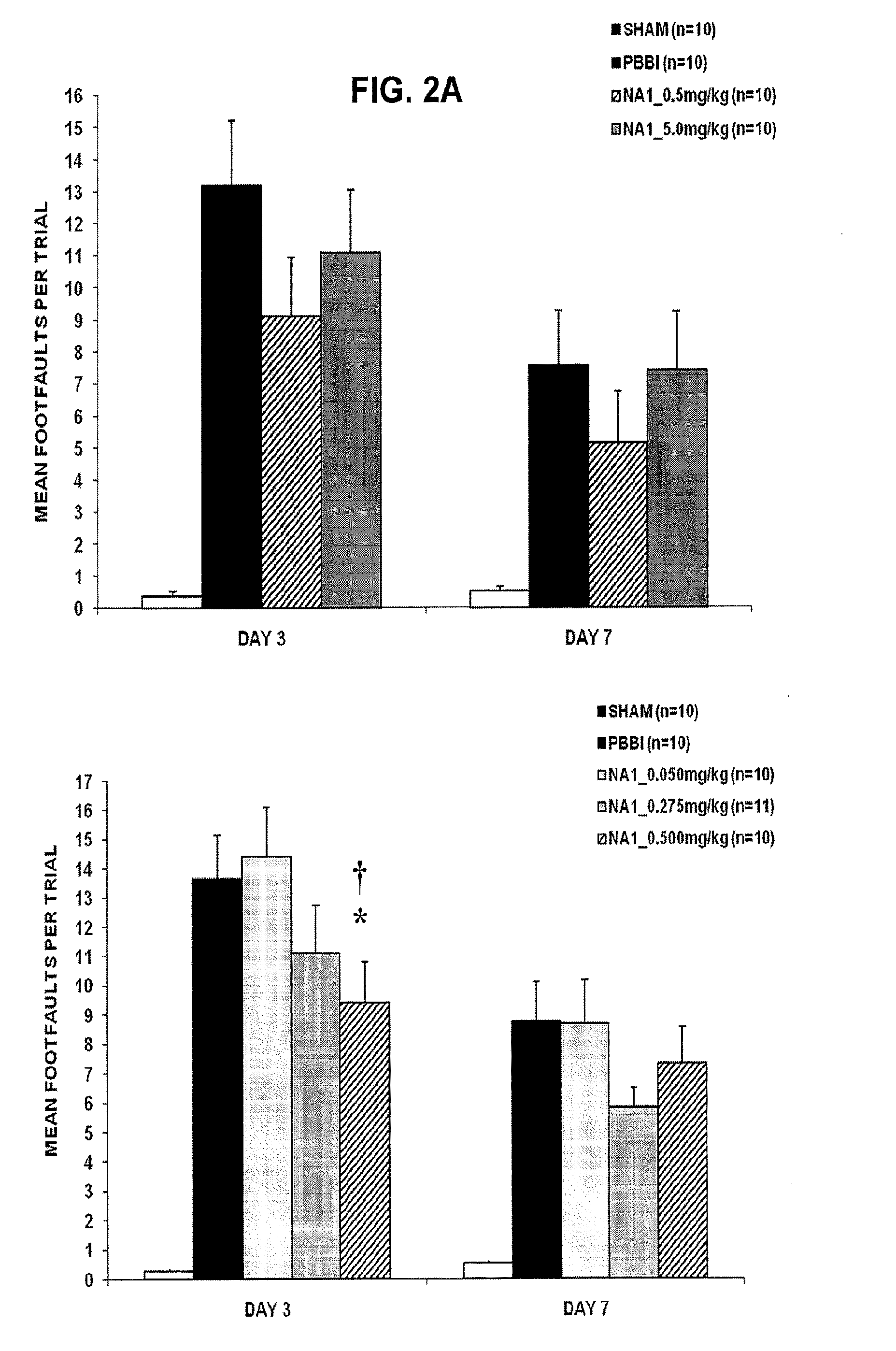
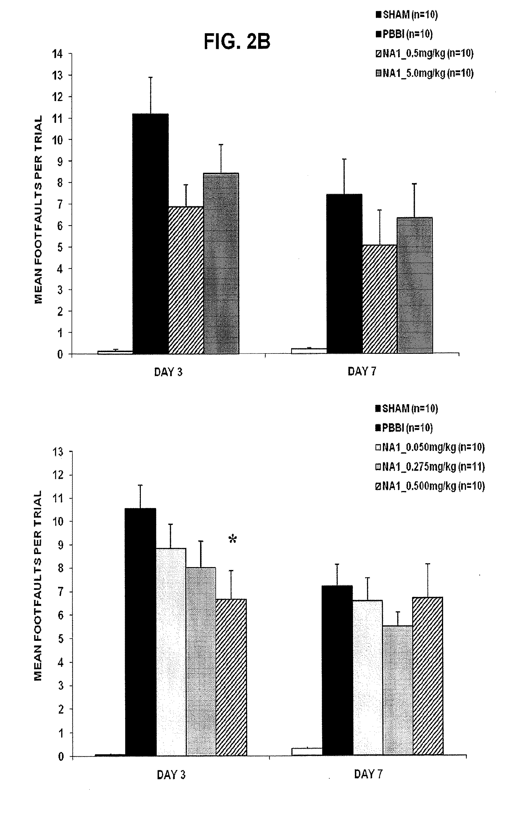
[0077]Male Sprague-Dawley rats (250-300 g) were subjected to a simulated ballistic wound to the right frontal hemisphere implemented by an inflatable penetrating probe. Three levels of injury severity were compared to control animals. Neurological and physiological outcome was assessed over a 3-day recovery period and brain tissue collected at 72 h for histopathological evaluation. Brain-injured regions included the ipsilateral frontal cortex and striatum with volumetric increases in intracranial hemorrhage (5-18 mm3) and lesion size (9-86 mm3) related to severity. Similarly, hemispheric swelling increased (3-14%) following PBI, associated with a significant rise in intracranial pressure. Astrogliosis was present in regions adjacent to the core-injury along with microglial and leukocyte infiltration. Injury remote to the lesion was observed in the cerebral peduncle that may have accounted, in part, for observed neurological deficits. Neurologi...
PUM
| Property | Measurement | Unit |
|---|---|---|
| concentration | aaaaa | aaaaa |
| concentration | aaaaa | aaaaa |
| temperature | aaaaa | aaaaa |
Abstract
Description
Claims
Application Information