Combined histological stain

a histological stain and fusion technology, applied in the field of histological sample visualization methods, can solve problems such as the inability to accurately quantify the staining

Active Publication Date: 2013-12-19
AGILENT TECH INC
View PDF4 Cites 7 Cited by
  • Summary
  • Abstract
  • Description
  • Claims
  • Application Information

AI Technical Summary

Benefits of technology

[0033]Advantageous features of the methods of the invention allow simultaneously visualizing the bulk of the target and single target entities (the same or different targets) of such as targets as proteins or cytoplasmic nucleic acids in one and the same histological sample stained with a histological stain. Use of a histological stain advantageously reveals characteristic morphological appearance of tissue of the sample for microscopic evaluation of the region

Problems solved by technology

the method provides a uniform staining pattern that appears to the microscopist as a homogeneous color with intracellular resolut

Method used


Image

  • Combined histological stain
  • Combined histological stain
  • Combined histological stain

Examples


examples

[0469]The below is a description of non-limiting selected working examples illustrating the invention.

Reagents

[0470]If not specified, the used reagents are either purchased from the recognized manufacturers or produced using the procedures described in WO2011047680 or WO2010094283 (the procedures are incorporated herein by reference) or follow the standard procedures of the art, e.g. procedures for solid-phase synthesis, conjugating polymers (including antibodies) with different labels, producing antibodies, antibody manipulation, etc. Exemplary production procedures for two selected compounds are described below.

Goat Anti-Rabbit Antibody Conjugated with Dex70 Conjugated with HRP (L348.111, Fractions 10-11.)

[0471]11 nmol 70 kDA MW dextran was reacted with 484 nmol HRP in 316 microliters of buffer A (100 mM NaCl, 25 mM NaHCO3, pH 9.5) for 3 h at 40 C. Thereafter 44 nmol Goat-anti-Rabbit 196 microL water was added to the dextran-HRP conjugate and allowed to react for further 1 h at 40...

experiment 1

Visualization of One Target by Staining (a) in a Histological Sample Stained with Haematoxylin and Eosin (HE Stain)

[0475]Slides with formalin fixed paraffin embedded sections of multiple human tissue samples were used as test material.

[0476]The slides were deparaffinated in xylene (2×5 min), 99% ethanol (2×2 min) then 70% ethanol. The slides were transferred to water for 5 min, then they were boiled in microwave oven in target retrieval solution for 10 min (Dako low pH, S1699).

[0477]Following cooling the slides were transferred to an Autostainer instrument and subjected to the following staining protocol:[0478]Pre rinse with wash buffer (Dako S3006)[0479]Peroxidase blocking solution (Dako S2023), 5 min.[0480]Wash (Dako S3006)[0481]Pan specific anti cytokeratin (Dako M3515) premixed with Goat-anti-Mouse-Dex150-HRP (L348.121), both 20 nM. then diluted to 20 pM, 5 min.[0482]Wash (Dako S3006)[0483]5 microM D21067, 0.28 mM DAB in incubation media 2, for 10 min.[0484]Wash (Dako S3006)[048...

experiment 2

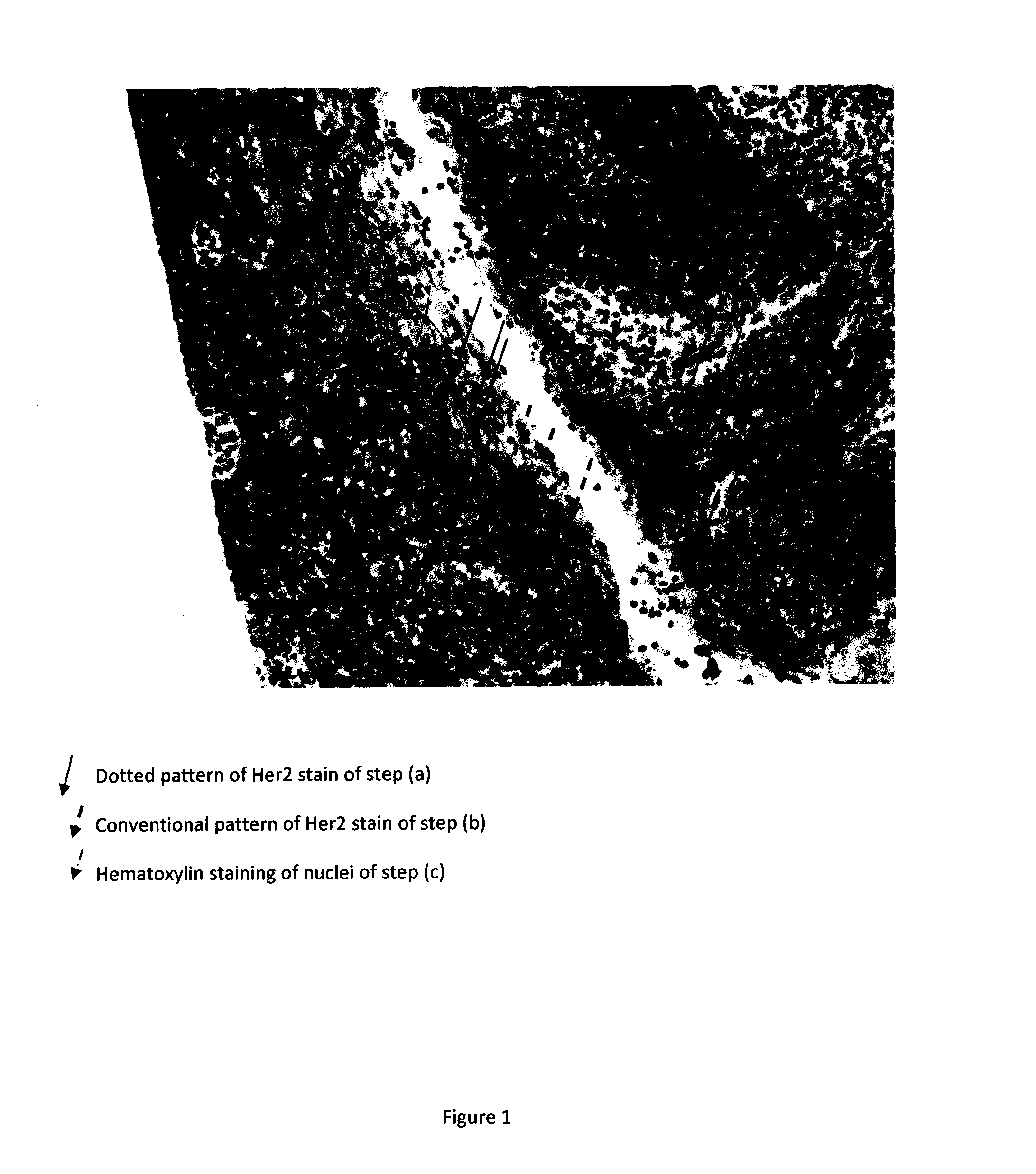
Visualization of One Target by Staining (a) and (b) in a Histological Sample Stained with Haematoxylin

[0498]Slides were pretreated as in experiment 1, and subjected to the following staining protocol on the Autostainer:[0499]Pre rinse with wash buffer (Dako S3006)[0500]Peroxidase blocking solution (Dako S2023), 5 min.[0501]Wash (Dako S3006)[0502]antiHer2 antibody, clone Dak 3-25-11, 6 nM for 20 min.[0503]Wash (Dako S3006)[0504]Goat-anti-Rabbit-Dextran70-HRP, (Lit 348.111, fraction 10-11) in incubation media 1. Varying concentration 12, 6, 3 and 1.5 picoM for 20 min.[0505]5 microM D21067, 0.28 mM DAB in incubation media 2, for 10 min.[0506]Wash (Dako S3006)[0507]antiFITC-F(ab)1-(AP)1, (D20036:), 20 nM in incubation media 1, 10 min[0508]Wash (Dako S3006)[0509]Liquid Permanent Red, (Dako K0640), 10 min[0510]Wash (Dako S3006)[0511]Goat-anti-Rabbit F(ab)1-(HRP), (D 20149), 40 nM in incubation media 1, 20 min

Or

[0512]Goat-anti-Rabbit F(ab)1-(HRP)1 (D 20149), 40 nM mixed with 40 nM unlabbel...


PUM

PropertyMeasurementUnit
Diameteraaaaaaaaaa
Diameteraaaaaaaaaa
Biological propertiesaaaaaaaaaa

Abstract

The present invention relates to methods of visualizing targets in histological samples, e.g. biopsy samples, wherein the methods comprise staining of the sample with (i) one or more target specific immunochemical stains, and (ii) a histological stain for specific tissue components e.g. iron, mucins glycogen, amyloid, nucleic acids, etc., e.g. hematoxilyn and/or eosin stains or the like, that is used to enhance contrast in the microscopic image of a tissue sample, highlight morphologic structures in the sample for viewing, define and examine tissues, cell populations, or organelles within individual cells. Methods may further comprise evaluation of expression of one or more targets in the sample. The disclosed methods are useful for medical diagnostics.

Description

FIELD OF INVENTION[0001]The present invention relates to methods of visualizing targets in histological samples, e.g. biopsy samples, wherein the methods comprise staining of the sample with one or more target directed immunochemical stains, and a histological stain for specific tissue components e.g. iron, mucins glycogen, amyloid, nucleic acids, etc., e.g. hematoxilyn and / or eosin stains or the like, that is used to enhance contrast in the microscopic image of a tissue sample, highlight morphologic structures in the sample for viewing, define and examine tissues, cell populations, or organelles within individual cells.BACKGROUND OF INVENTION[0002]Histopathology, the microscopic study of diseased tissue, is an important tool in anatomical pathology, since accurate diagnosis of cancer and other diseases usually requires histopathological examination of samples.[0003]Analysis of the microscopic anatomy of a sample of patient tissue is usually performed by examining under a light micr...

Claims


Application Information

Patent Timeline
IPC IPC(8): G01N33/58
CPCG01N33/581G01N33/52G01N33/53G01N33/86
Inventor LOHSE, JESPER
Owner AGILENT TECH INC
Why Eureka
Try Eureka