Enzymatic metal nanoparticle sensor for detecting DNA binders
a metal nanoparticle and sensor technology, applied in the field of biochemistry and nanotechnology, can solve the problems of multiple tedious steps or stringent assay conditions, incubation period, false positive, etc., and achieve the effect of low specificity, fast, simple and specific detection methods
- Summary
- Abstract
- Description
- Claims
- Application Information
AI Technical Summary
Benefits of technology
Problems solved by technology
Method used
Image
Examples
example 1
Aggregation Kinetics of dsDNA-Functionalized AuNPs in the Presence of DNase I
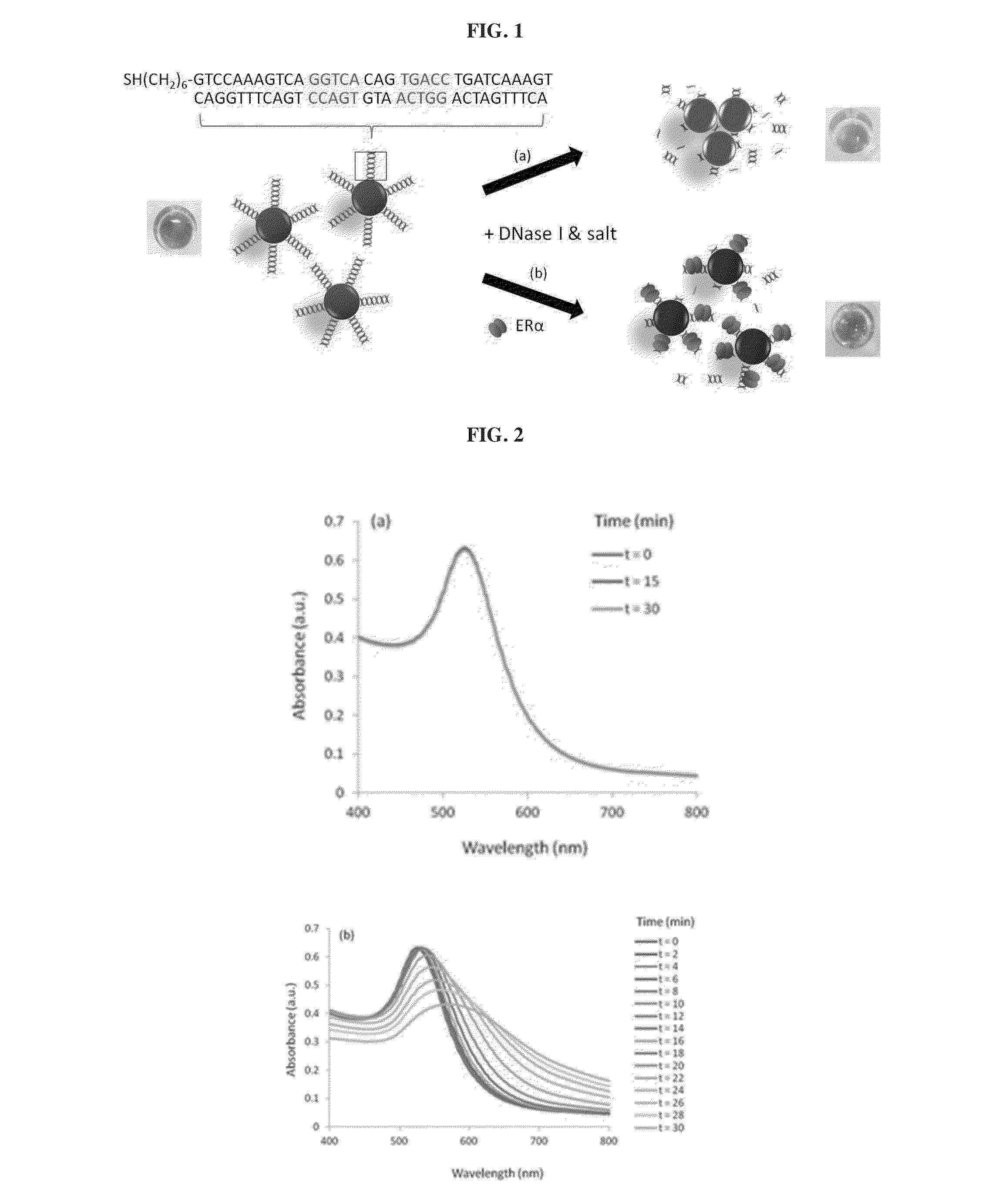
[0117]An application is described herein for detecting estrogen receptor (ER)-DNA interaction. ER is a DNA-binding protein that binds specifically to a dsDNA sequence known as estrogen-responsive elements (ERE), to regulate target gene transcription.
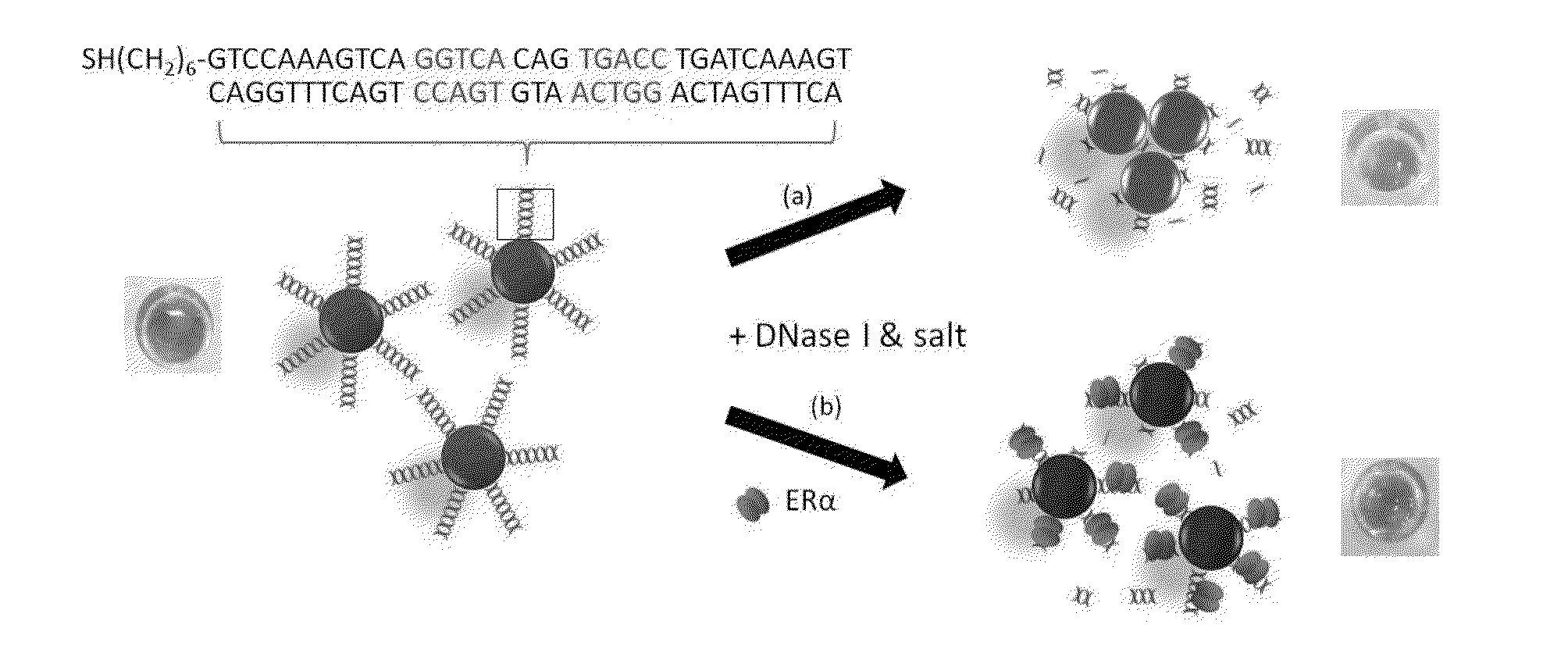
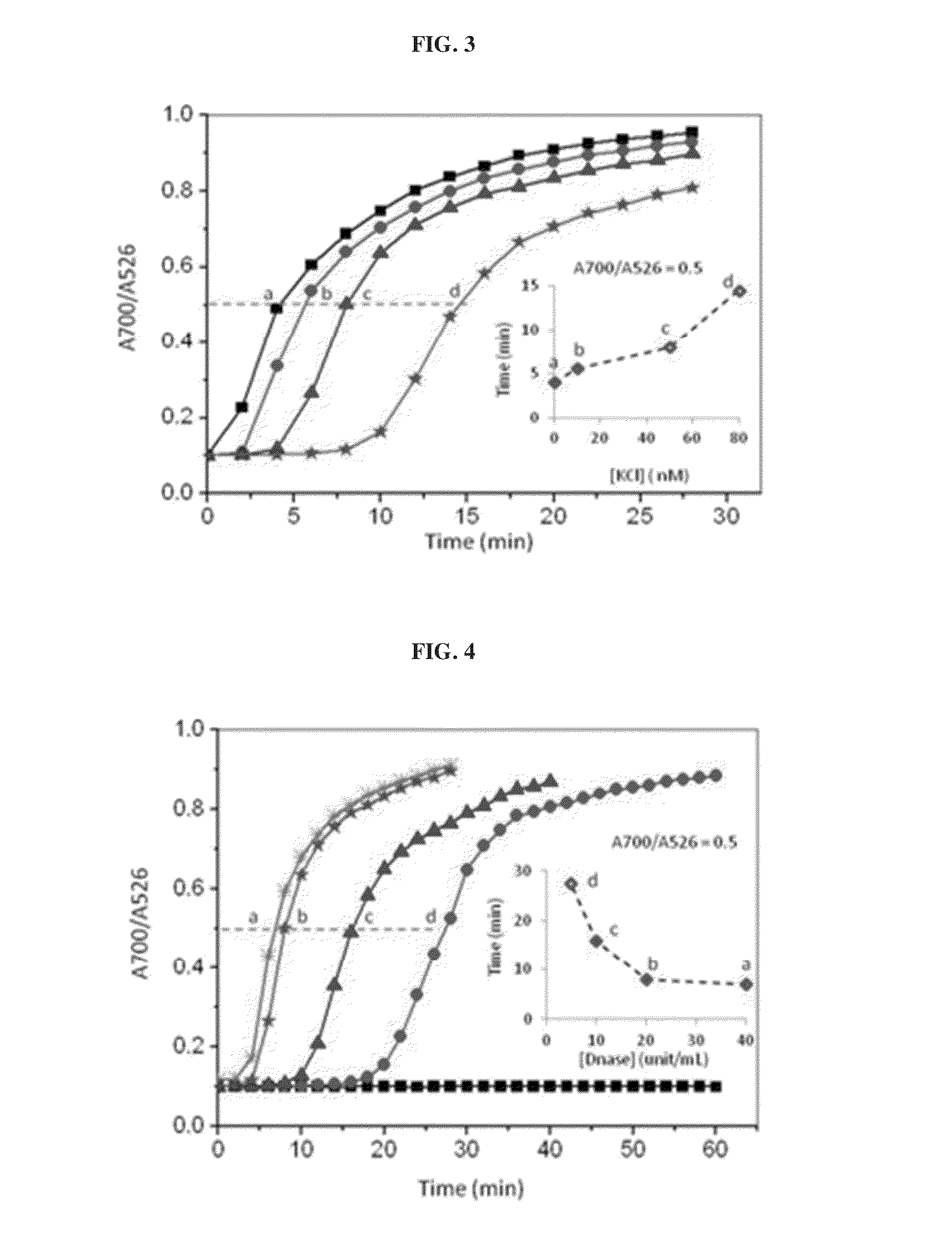
[0118]In this example, DNase I is used to cleave the DNA conjugated on the AuNPs surface to induce particle aggregation by the MgCl2 and KCl content of the DNase I buffer. Double-stranded (ds) ERE sequences from the vitellogenin A2 gene (vit) containing the core binding sequence of 5′-GGTCAnnnTGACC-3′ (SEQ ID No. 1; n: spacer nucleotides) of ERα is conjugated on the AuNPs following the previously reported protocol. As shown in FIG. 2a, the as-conjugated dsvit-AuNPs is stable in DNase I buffer solution (20 mM MgCl2 and 50 mM KCl) for at least 30 mM. When 5 unit / mL of DNase was added to the dsVit-AuNP in the same buffer solution, aggregation of AuNPs was observed,...
example 2
Detection of ERα Using dsDNA-Modified AuNPs and DNase
[0121]In the example 1 above, it has been shown that the aggregation of AuNPs resulting from the enzymatic cleavage of the DNA conjugated to the particle surface can be detected immediately by naked eyes observation of color changes and / or quantified by the absorption spectra measurement. Using the ERα and their DNA response elements as a model system (FIG. 5a), the inventors demonstrated that the presence of ERα and its binding to the specific DNA on AuNPs can be detected based on the retarded / absent DNA cleavage, which in turn keeps the nanoparticles dispersed (and thus red) under high salt conditions, while the sample without ERα experienced extensive aggregation (purple) in the same salt and DNase I concentration. To examine the effects of the ERα protection on AuNPs, concentration of ERα was varied with a fixed amount of DNase I (20 units / mL). FIG. 5b shows that decreasing concentration of ERα in the sample results in an incr...
example 3
Detection of ERα in Nuclear Extracts from MCF-7 Human Breast Cancer Cell
[0123]Nuclear extracts contain thousands of different transcription factors. It thus renders detection and identification of a DNA binding proteins of interest, particularly challenging in terms of selectivity and specificity. Sure enough, the method should allow specific binding of one particular proteins among a mixture of thousand others different DNA binding proteins in a physiological setting (i.e. the nucleus of isolated cells)
[0124]The inventors used the DNase-assisted AuNPs assay to detect the binding of native (i.e. as opposed to recombinant or ERα from human MCF-7 breast cancer cells extracts to its DNA response element. It was found that the usage of dsvit-AuNPs alone failed to detect the ERα-DNA binding (FIG. 7a) upon addition of DNase, due to insufficient amount of native ERα expression in the biological samples. However, the MCF (ER+) sample is differentiable from the MCF (ER−) sample when 30 nM of...
PUM
| Property | Measurement | Unit |
|---|---|---|
| size | aaaaa | aaaaa |
| size | aaaaa | aaaaa |
| size | aaaaa | aaaaa |
Abstract
Description
Claims
Application Information
 - R&D
- Intellectual Property
- Life Sciences
- Materials
- Tech Scout
- Unparalleled Data Quality
- Higher Quality Content
- 60% Fewer Hallucinations
Browse by: Latest US Patents, China's latest patents, Technical Efficacy Thesaurus, Application Domain, Technology Topic, Popular Technical Reports.
© 2025 PatSnap. All rights reserved.Legal|Privacy policy|Modern Slavery Act Transparency Statement|Sitemap|About US| Contact US: help@patsnap.com


