
Method of diagnosing neoplasms

a neoplasm and neoplasm technology, applied in the field of neoplasm diagnosis, can solve the problems of blood loss, unpredictability of current or future risk of cancer, and observation error and confusion of all those other than number and siz

Inactive Publication Date: 2014-06-05
CLINICAL GENOMICS PTY LTD +1
View PDF0 Cites 4 Cited by
  • Summary
  • Abstract
  • Description
  • Claims
  • Application Information

AI Technical Summary

Benefits of technology

The present invention provides a method for screening for the onset or predisposition to the onset of a large intestine neoplasm in an individual by measuring the expression levels of certain genes or regions of chromosomes. This method can help identify individuals at risk for colon cancer and other gastrointestinal cancers, which can aid in the early detection and treatment of these diseases.

Problems solved by technology

However, this is related to size of an adenoma.
Except for the presence of adenomas and its size, none of these is objectively defined and all those other than number and size are subject to observer error and to confusion as to precise definition of the feature in question.
Because such factors can be difficult to assess and define, their value as predictors of current or future risk for cancer is imprecise.
Colonic adenomas are localised areas of dysplastic epithelium which initially involve just one or several crypts and may not protrude from the surface, but with increased growth in size, usually resulting from an imbalance in proliferation and / or apoptosis, they may protrude.
All of these descriptors, with the exception of number and size, are relatively subjective and subject to interobserver disagreement.
None except size or number is objective and all are relatively subjective and subject to interobserver disagreement.
Adenomas are generally asymptomatic, therefore rendering difficult their diagnosis and treatment at a stage prior to when they might develop invasive characteristics and so became cancer.
Some adenomas result in blood loss which might be observed or detectable in the stools; while sometimes visible by eye, it is often, when it occurs, microscopic or “occult”.
However, since blood in the stool, whether overt or occult, can also be indicative of non-adenomatous conditions, the accurate diagnosis of adenoma is rendered difficult without the application of highly invasive procedures such as colonoscopy combined with tissue acquisition by either removal (i.e. polypectomy) or biopsy and subsequent histopathological analysis.
These adenomas may be high risk, advanced or neither of these.
Furthermore, it can be difficult after colonoscopy, to be certain that all adenomas have been removed, especially in a person who has had multiple adenomas.
However, from the patient perspective, harvesting colorectal tissue samples is invasive and not without risk in terms of post-operative complications, such as infection.
In work leading up to the present invention it has been determined that in the context of colorectal neoplasia gene markers, the level of expression of which are increased in neoplasia, this increase in expression is not necessarily easily detectable in the plasma due to issues of diagnostic sensitivity.
However, it has been unexpectedly found that a small subset of these gene markers exhibit a significant increase in the level of their expression within circulating exosomes.

Method used


Image

  • Method of diagnosing neoplasms

Examples


example 1

[0144]To test the detectability of gene markers in blood plasma specimens, commercially available TaqMan assays were purchased from Applied Biosystems. Positioning of PCR amplicon locations (ie which exon-exon junction) was guided by the Human ST Exon 1.0 microarray study of 42 matched normal specimens and serrated adenomas, ie. Towards exons showing the highest fold difference between normal and adenomous colon specimens.

[0145]A total of 68 TaqMan assays targeting a total of 46 genes (red / green coloured genes in Appendix 1 and 2) were used on 2.5 uL cDNA generated from RNA (RNA:cDNA 1:1) extracted from 2 mL plasma. Plasma was produced from two consecutive centrifugation steps (1,500 g, 10 min, 4 deg C) of whole blood collected in 9 mL K3-EDTA vacutainer blood tubes. The TaqMan assays were tested on a least one panel of 45 blood plasma specimens from 15 normal patient, 15 patients with colorectal adenomas and 15 patients with colorectal cancer (phenotypes obtained by colonoscopy). T...

example 2

Materials and Methods

Clinical Specimens

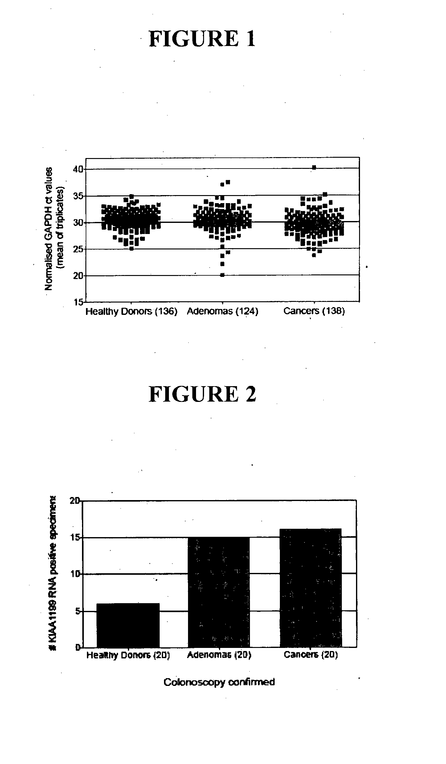
[0149]Blood specimens from healthy donors (136), adenoma (124, any grade), and cancer (138, any grade) patients were procured through collaboration with Flinders Medical Center (Adelaide, Australia) or from a clinical specimen vendor (Proteogenex, USA). Colorectal neoplastic status was confirmed by colonoscopy and pathology review for all specimens. Plasma was generated from whole blood phlebotomy specimens (K3EDTA Vacutainer) within 4 hrs of blood draw using a 2×1,500 g spin protocol.

Plasma RNA Extraction, Generation of cDNA Libraries and Extraction Quality Control

[0150]RNA was extracted from 2 mL plasma aliquots using the QIAamp Circulating Nucleic Acid Extraction Kit (Qiagen, Australia) and eluted into a final volume of 100pL. To normalize for nucleic acid extraction efficiency differences between specimens, arRNA enterovirus (Asuragen, US) was spiked into each plasma specimen prior to RNA isolation and recovery was measured downstream of th...


PUM

PropertyMeasurementUnit
sizeaaaaaaaaaa
sizeaaaaaaaaaa
volumeaaaaaaaaaa

Abstract

The present invention relates generally to a method for screening a subject for the onset, predisposition to the onset and / or progression of a colorectal neoplasm by screening for modulation in the level of expression of one or more nucleic acid markers. More particularly, the present invention provides a method for screening a subject for the onset, predisposition to the onset and / or progression of a colorectal neoplasm by screening for modulation in the level of expression of one or more gene markers in membranous microvesicles. The expression profiles of the present invention are useful in a range of applications including, but not limited to, those relating to the diagnosing and / or monitoring of colorectal neoplasms, such as colorectal adenoma and adenocarcinomas.

Description

FIELD OF THE INVENTION[0001]The present invention relates generally to a method for screening a subject for the onset, predisposition to the onset and / or progression of a colorectal neoplasm by screening for modulation in the level of expression of one or more nucleic acid markers. More particularly, the present invention provides a method for screening a subject for the onset, predisposition to the onset and / or progression of a colorectal neoplasm by screening for modulation in the level of expression of one or more gene markers in membranous microvesicles. The expression profiles of the present invention are useful in a range of applications including, but not limited to, those relating to the diagnosing and / or monitoring of colorectal neoplasms, such as colorectal adenoma and adenocarcinomas.BACKGROUND OF THE INVENTION[0002]Bibliographic details of the publications referred to by author in this specification are collected alphabetically at the end of the description.[0003]The ref...

Claims


Application Information

Patent Timeline
Patent Type & Authority Applications(United States)
IPC IPC(8): C12Q1/68
CPCC12Q1/6886G01N33/57419G01N2800/56G01N2800/52G01N2800/50C12Q2600/158G01N33/574
Inventor LAPOINTE, LAWRENCE CHARLESPEDERSEN, SUSANNE KARTINMOLLOY, PETER
Owner CLINICAL GENOMICS PTY LTD
Learn More