Image analysis device, image analysis method, and image analysis programme

a technology of image analysis and image analysis, applied in the direction of image enhancement, measurement using nmr, instruments, etc., can solve the problems of limited image resolution, limitation of imaging time, and inability to directly portray anmyloid by means of current image detecting device for health care services, etc., to achieve stable verification, short imaging time, and low magnetic field intensity

Active Publication Date: 2014-08-21
NAT UNIV CORP KUMAMOTO UNIV
View PDF1 Cites 2 Cited by
  • Summary
  • Abstract
  • Description
  • Claims
  • Application Information

AI Technical Summary

Benefits of technology

[0012]As described above, according to one or more embodiments of the present invention, a minute change in tissue may be detected by using an MRI phase difference image which is sensitive to a minute magnetic change of the tissue. In other words, a phase change due to a partial volume effect of the tissue is determined as a change in phase difference distribution of a predetermined target area (in one or more embodiments below, referred to as an area of interest), and as a result, existence of the target tissue may be stably verified by using an MRI which has a short imaging time and low magnetic field intensity.
[0013]Instead of directly creating an image of pixel by pixel having high accuracy by an unrealistic method for a long imaging time, it is considered that a detecting method using short-time imaging or low-magnetic field equipment is facilitated in the industry and the medical field. Further, the method may be used without changing a device in the related art or introducing an additional device because of software processing.

Problems solved by technology

However, a diameter of the amyloid β is about 0.1 mm or less, and it is considered that the amyloid β cannot directly be portrayed by means of a current image detecting device for health care services.
However, in the medical field, there is a limitation of an imaging time, and there is a limitation of the resolution in the image which can be imaged within the limited time.
Of course, in high-resolution image equipment, the portraying is possible, but the high-resolution image equipment is beyond a range of the health care services, and expensive.
An image diagnosis method requiring expensive equipment which is beyond the range of the health care services provided under medical insurance cannot be said as the solution of a current urgent medical problem, and a method of detecting a tissue or lesions, which cannot be portrayed by means of medical imaging equipment such as an MRI, realistically within a current range of health care services provided under medical insurance has been required.

Method used


Image

  • Image analysis device, image analysis method, and image analysis programme
  • Image analysis device, image analysis method, and image analysis programme
  • Image analysis device, image analysis method, and image analysis programme

Examples


Embodiment Construction

[0024]In embodiments of the invention, numerous specific details are set forth in order to provide a more thorough understanding of the invention. However, it will be apparent to one with ordinary skill in the art that the invention may be practiced without these specific details. In other instances, well-known features have not been described in detail to avoid obscuring the invention.

[0025]Hereinafter, embodiments of the present invention will be described according to the following order.

[0026](1) Configuration of embodiments:

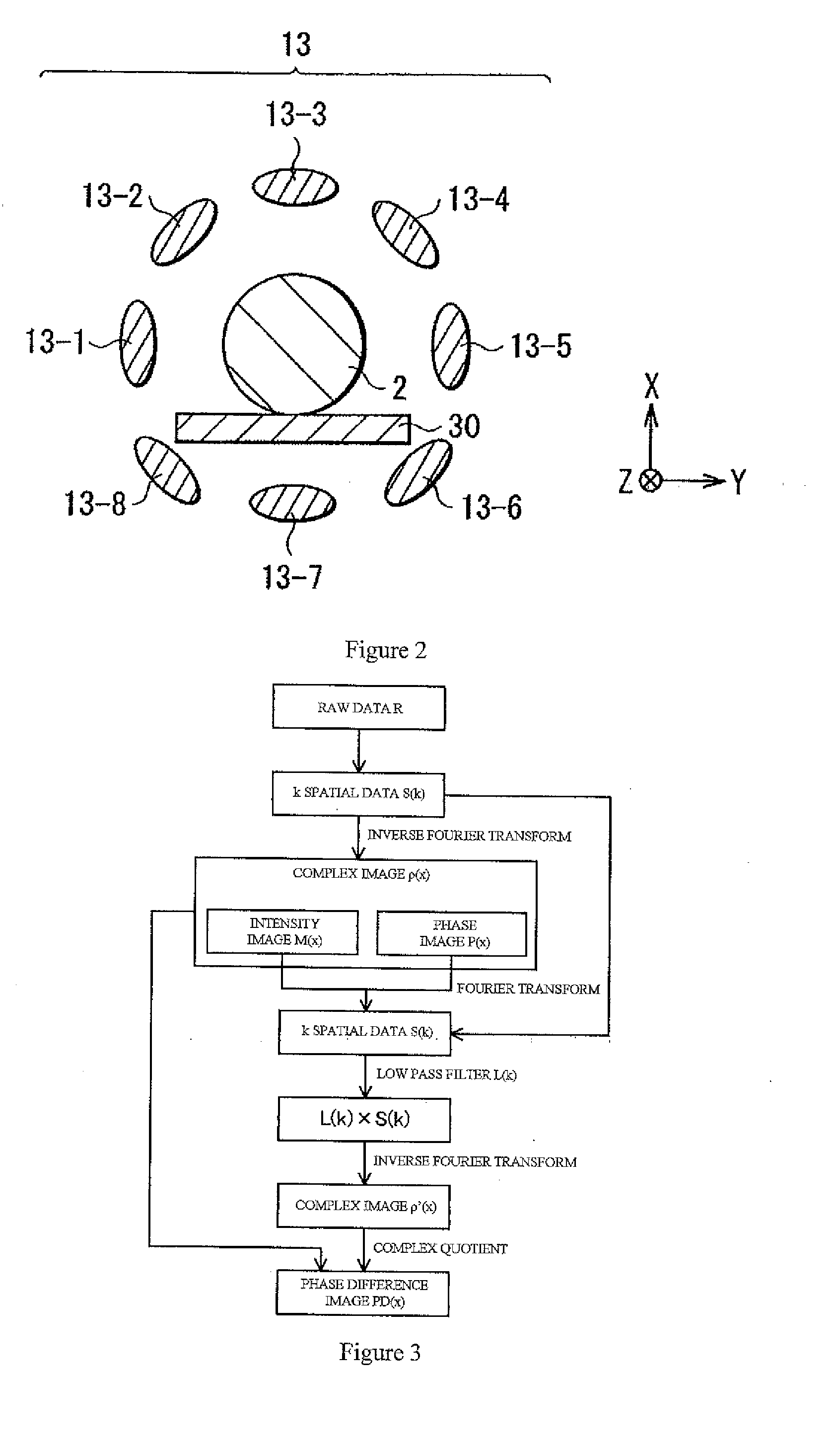
[0027](2) Creating phase difference image:

[0028](3) Determining process:

[0029](4) Creating of shape image:

[0030](5) Summary:

(1) Configuration of Embodiments

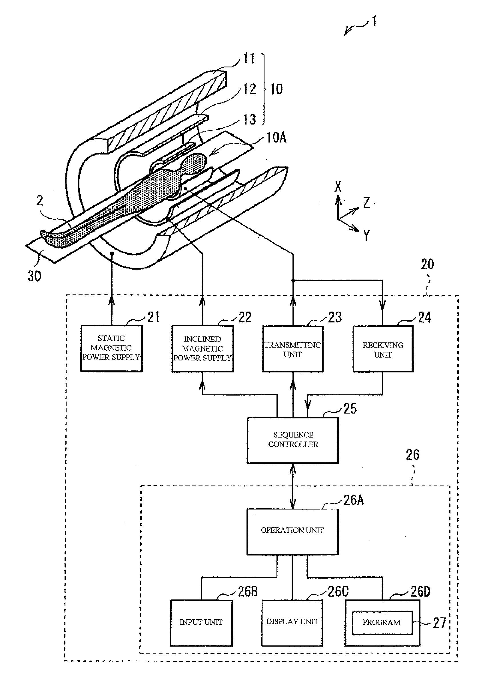
[0031]FIG. 1 is a diagram illustrating a schematic configuration of a magnetic resonance imaging (MRI) device 1. The MRI device 1 is a device of imaging internal information of a subject 2 by using an NMR phenomenon. The MRI device 1 is a new type MRI device that portrays a shape image by using a phase im...


PUM


Abstract

An image analysis device for analyzing a magnetic resonance image obtained from a living body includes a phase difference distribution creating unit configured to create a phase difference distribution of a magnetic resonance image obtained from a predetermined area of the living body, a fitting unit configured to fit the phase difference distribution created by the phase difference distribution creating unit with a plurality of function groups, and a verifying unit configured to verify normality of the living body included in the predetermined area, based on the magnetic susceptibility of the tissue included in the predetermined area determined on the basis of the parameters of the plurality of function groups fit to the phase difference distribution by the fitting unit.

Description

TECHNICAL FIELD [0001]The present invention relates to an image analysis device, an image analysis method, and an image analysis program, and more particularly, to an image analysis device, an image analysis method, and an image analysis program for analyzing a magnetic resonance image obtained from a living body.BACKGROUND ART [0002]An Alzheimer's disease which is one of dementia is the current major problem, and has been studied in the world without exception. One of main causes of the Alzheimer's disease is known as a protein called amyloid β accumulated in the brain. However, a diameter of the amyloid β is about 0.1 mm or less, and it is considered that the amyloid β cannot directly be portrayed by means of a current image detecting device for health care services.[0003]By the way, most of medical imaging equipment such as an MRI is aimed at directly portraying a target tissue or lesions. However, in the medical field, there is a limitation of an imaging time, and there is a lim...

Claims


Application Information

Patent Timeline
Patent Type & Authority Applications(United States)
IPC IPC(8): G06T7/00
CPCG06T2207/30004G06T7/0012A61B5/055A61B5/4088G01R33/5608G01R33/24G01R33/56
Inventor YONEDA, TETSUYA
Owner NAT UNIV CORP KUMAMOTO UNIV
Why Eureka
Try Eureka