

Photosensitive cardiac rhythm modulation systems
a technology of cardiac rhythm and modulation system, applied in the field of photosensitive cardiac rhythm modulation system, can solve the problems of increasing the patient's inflammatory response and risk of infection, limited battery life, and unresponsive implantable devices to autonomic heart rate modulation, so as to suppress the generation of action potential and suppress the effect of beating
- Summary
- Abstract
- Description
- Claims
- Application Information
AI Technical Summary
Benefits of technology
Problems solved by technology
Method used
Image

Examples
example 1
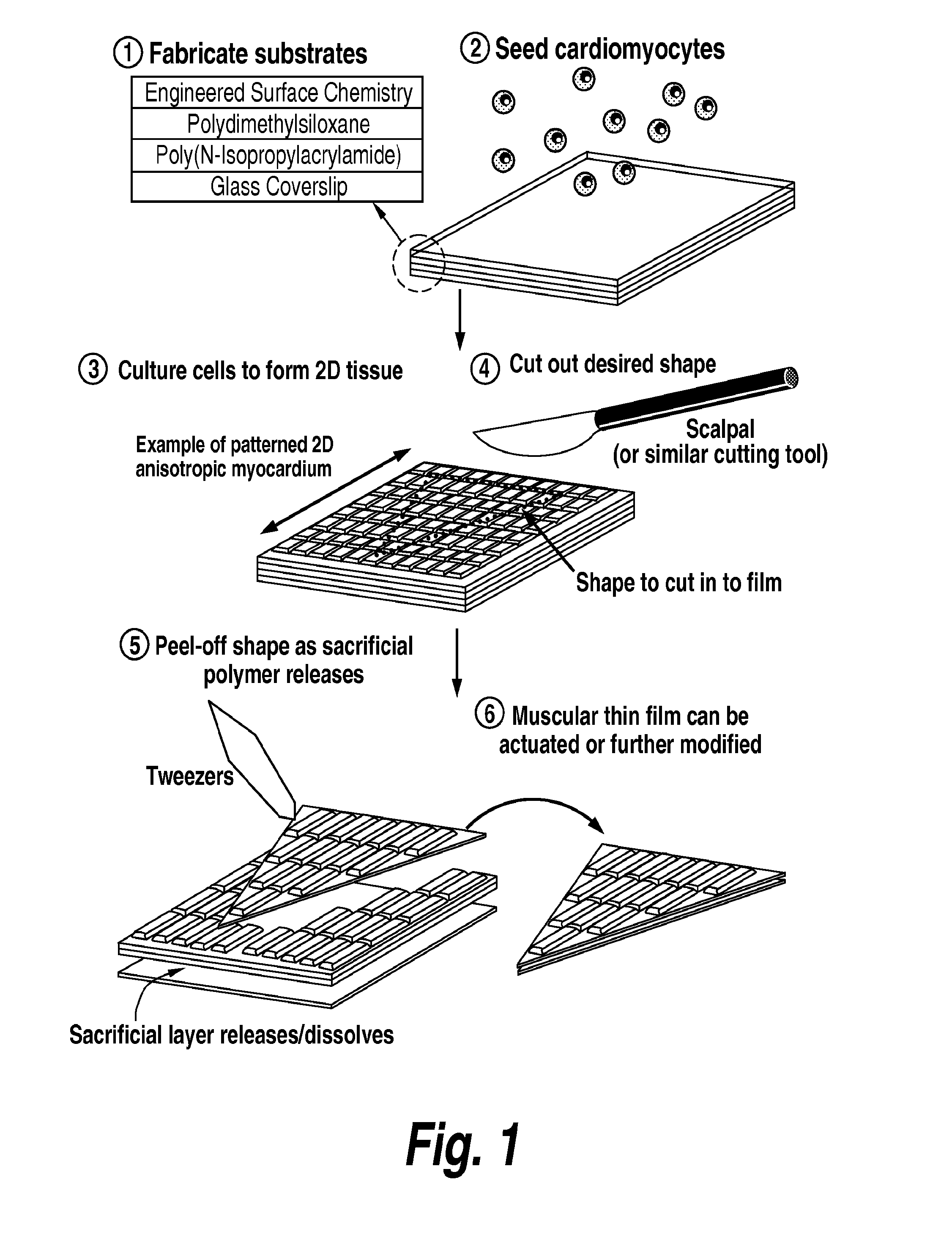
Construction of Pacing Muscular Thin Films
[0102]Tissue engineered pacemakers are made by harvesting the sinoatrial node from neonatal rat right atria, chemically dissociating the cells, and culturing them on micropatterned MTFs. More specifically, the right atria from neonate rats is harvested, carefully dissected, and those myocytes in the region of the sinoatrial node are chemically dissociated. These myocytes are cultured on micropatterned MTFs to form an anisotropic tissue structure with autonomous beating capability.
[0103]To demonstrate that the tissue engineered pacemaker is capable of integration into and pacing control of cardiac tissue in vitro, immunohistochemical analysis is performed of a pacing MTF placed on and attached to engineered ventricular myocardium (FIG. 5). Staining is used to demonstrate the formation of gap junctions between the ventricular myocardium and the pacemaking cells. It has been shown that ventricular myocytes predominantly express C×43 gap junctio...
example 2
Surgical Implantation of Pacing Muscular Thin Films in Rats
[0105]Surgical implantation of pacing MTFs is accomplished by surgically ablating the sinoatrial node in anaesthetized rats and sewing pacing MTFs on the apical surface of the right atria. More specifically, surgical ablation of the sinoatrial node is accomplished by cauterizing the node in vivo during survival surgery (Tarnayski et al., Physiol. Genomics: 16: pp. 349-360, 2004). Pacing is restored by implantation of a pacing MTF constructed as described in Example 1. Briefly, 70 mg / kg of pentobarbital sodium is administered to induce anesthesia. After an adequate depth of anesthesia is attained, the rat is placed in a supine position and a taut 5-0 ligature is situated behind the front upper incisors to keep the neck slightly extended. The tongue is retracted and held with forceps while inserting a 20 gauge catheter into the trachea. The catheter is then attached to a ventilator via a Y-shaped connector. Ventilation is perf...
example 3
Generation of Photoinduced Action Potentials in 2-Dimensional Genetically Engineered Photosensitive Cardiac Tissue
[0107]Isotropic and anisotropic 2-dimensional genetically engineered photosensitive cardiac tissues are prepared as described herein. In general, one or more of the cells used to prepare the 2-dimensional genetically engineered photosensitive cardiac tissue is genetically modified to express a photosensitive membrane transport mechanism for the purpose of generating photoinduced action potentials. Genetic modification is carried out via viral or non-viral transfection. Alternatively, tissues fabricated from cells derived from a cell line which stably expresses a photosensitive membrane transport mechanism may be used to prepare isotropic and anisotropic 2-dimensional genetically engineered photosensitive cardiac tissues. Suitable cells for preparation of these tissues include, but are not limited to, primary mammalian cardiac myocytes, cardiac cells derived from embryoni...
PUM
| Property | Measurement | Unit |
|---|---|---|
| Action potential | aaaaa | aaaaa |
| Flexibility | aaaaa | aaaaa |
| Wavelength | aaaaa | aaaaa |
Abstract
Description
Claims
Application Information